Review
. 2010 Jul;14(7):1917-21.
doi: 10.1111/j.1582-4934.2010.01111.x. Epub 2010 Jul 5.

Myocardial telocytes: a specific new cellular entity

Affiliations
Review

Myocardial telocytes: a specific new cellular entity

Sawa Kostin. J Cell Mol Med. 2010 Jul.

Abstract

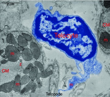
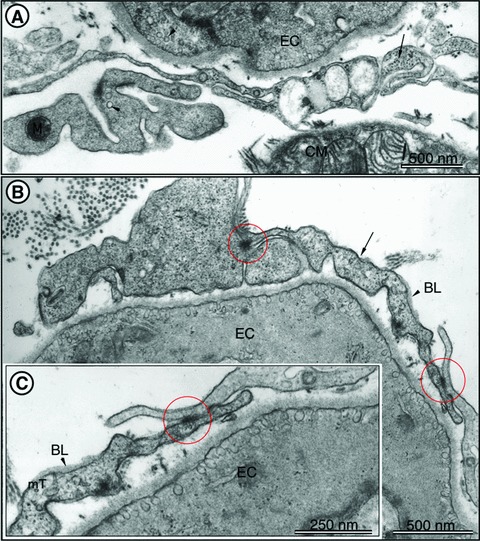
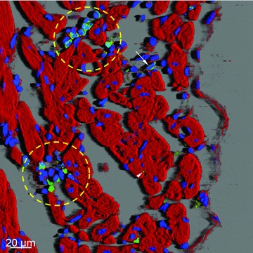
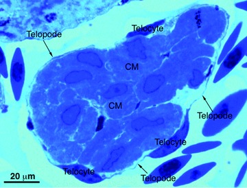
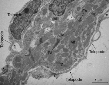
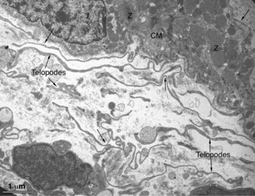
The existence of a new type of interstitial cells in the heart namely, interstitial Cajal-like cells (ICLC), has been described for the first time by Hinescu and Popescu in 2005. This study was then followed by an ascending trend of publications regarding the morphology, phenotype and distribution of myocardial ICLC in diverse species including human patients. Recently the new term 'telocytes' has been proposed for cells formerly known as ICLC, and the term 'telopodes' has been proposed for the prolongations of these cells. The identification of these cells is based on ultrastructural criteria. In addition, telocyters/telyopodes can be identified by several complementary approaches including methylene blue vital staining, silver impregnation and immunoreactivity against CD117/c-kit, vimentin, etc. This point of view presents critical data existing in literature, as well as own results, which unequivocally provide compelling evidence that telocytes are a new distinct cellular entity of myocardial interstitium. Several presumable functions of the myocardial telocytes are discussed: (i) intercellular signalling, (ii) cardiac repair/remodelling and (iii) stem cell nursing in cardiac renewal.


Figures

Fig 1
Fig 1
Digitally coloured electron micrograph of rat ventricular myocardium showing a typical cell body (telocyte, blue) in between two cardiomyocytes (CM). Note the sharp transition (Arrow) from cell body to long tortuous process (telocytes). m – mitochondria, Z – Z-band. Reproduced with permission from [3].
Fig 2
Fig 2
TEM images of rat left ventricular myocardium. (A) A typical example of telopodes (arrows) forming a labyrinthine system and located in vicinity of endothelial cells (EC) and cardiomyocytes. The presence of caveolae (arrowheads) is a typical feature of telopodes and ECs. Note the cross-section of intermediate filaments (arrow) inside the telopodes. (B) Telopodes (arrows) located in close vicinity of EC. Cell-to-cell contacts of telopodes are indicated with circles. Note the presence of the basal lamina (BL, arrowhead), which can occasionally be observed in telopodes. (C) Higher magnification of the right part of (B) showing junctional complexes (circle) between two telopodes. BL – basal lamina. Cross-sectioned microtubules (mT) can be seen in telopodes. Reproduced with permission from [3].
Fig 3
Fig 3
Confocal microscopy of c-kit+ cells in human left ventricular myocardium. Three-dimensional shadow projection image of transversally and obliquely oriented fibres image showing niche-like clusters (yellow round marks) or isolated round c-kit+ cells (arrow) in the subepicardial loose connective tissue. Note small green dots (arrowhead) which represent transversally sectioned telopodes of c-kit+ cells located in close proximity to cardiomyocytes. F-actin is red stained with phalloidin conjugated with TRITC and nuclei are blue stained with DAPI.
Fig 4
Fig 4
Light microscopy of toluidine-blue stained semithin sections of the normal heart of the newt ‘Notophthalmus viridescens’. A typical trabecula containing cardiomyocytes (CM) which are completely circumscribed by numerous telocytes and very thin telopodes (arrows).
Fig 5
Fig 5
Transmission electron microscopy of the normal zebrafish heart. A typical trabecula containing cardiomyocytes (CM) which are in close contact with telocytes and telopodes (arrows). Asterisk indicates a dichotomous pattern of telocytes. M – mitochondria, Mf – myofilaments.
Fig 6
Fig 6
Transmission electron microscopy of the injured heart of the newt Notophthalmus viridescens. Numerous telopodes (arrows) are present in a healing myocardial region 4 days hours after amputation of the ventricular apex. Telopodes seem to originate from the intact myocardium and to extend to the injured site and show a strong tendency to form a cellular network.

References

    1. Popescu LM, Faussone-Pellegrini MS. TELOCYTES – a case of serendipity: the winding way from interstitial cells of Cajal (ICC), via interstitial Cajal-like cells (ICLC) to TELOCYTES. J Cell Mol Med. 2010;14:729–40. - PMC - PubMed
    1. Popescu LM, Gherghiceanu M, Hinescu ME, et al. Insights into the interstitium of ventricular myocardium: interstitial Cajal-like cells (ICLC) J Cell Mol Med. 2006;10:429–58. - PMC - PubMed
    1. Kostin S, Popescu LM. A distinct type of cell in myocardium: interstitial Cajal-like cells (ICLCs) J Cell Mol Med. 2009;13:295–308. - PMC - PubMed
    1. Hinescu ME, Gherghiceanu M, Mandache E, et al. Interstitial Cajal-like cells (ICLC) in atrial myocardium: ultrastructural and immunohistochemical characterization. J Cell Mol Med. 2006;10:243–57. - PMC - PubMed
    1. Popescu L, Gherghiceanu M, Manole C, Faussone-Pellegrini MS. Cardiac renewing: interstitial Cajal-like cells nurse cardiomyocyte progenitors in epicardial stem cell niches. J Cell Mol Med. 2009;13:866–86. - PMC - PubMed