

Hemolysis and acute kidney failure
- PMID: 20605299
- PMCID: PMC3282484
- DOI: 10.1053/j.ajkd.2010.03.025
Hemolysis and acute kidney failure
Abstract
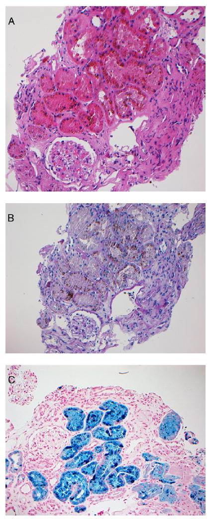
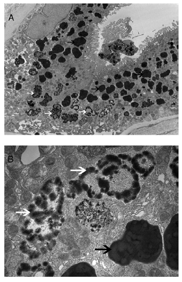
Deposits of iron and hemosiderosis in the kidney have been observed in diseases with intravascular hemolysis, including paroxysmal nocturnal hemoglobinuria, and valvular heart diseases and prosthetic heart valve implants. However, the decrease in kidney function associated with hemolysis caused by cardiac valvular disease or prostheses is less well recognized. We present a case of intravascular hemolysis after repair and banding of the mitral valve that resulted in massive renal tubular deposition of hemosiderin with decreased kidney function. We discuss the pathophysiologic process of both acute and chronic tubular injury from heme and heme proteins, including injury to organelles resulting in autophagic vacuoles containing damaged organelles, such as mitochondria. We conclude that tubular injury resulting from heme proteins should be considered as a cause of decreased kidney function in all patients with a cardiac valvular disease or prosthesis.
Copyright © 2010 National Kidney Foundation, Inc. Published by Elsevier Inc. All rights reserved.
Conflict of interest statement
Figures
References
-
- Leonardi P, Ruol A. Renal hemosiderosis in the hemolytic anemias: diagnosis by means of needle biopsy. Blood. 1960;16:1029–1038. - PubMed
-
- Nath KA, Vercellotti GM, Grande JP, et al. Heme proteininduced chronic renal inflammation: suppressive effect of induced heme oxygenase-1. Kidney Int. 2001;59(1):106–117. - PubMed
-
- Roberts WC, Morrow AG. Renal hemosiderosis in patients with prosthetic aortic valves. Circulation. 1966;33(3):390–398. - PubMed
-
- Ackermann D, Vogt B, Gugger M, Marti HP. Renal haemosiderosis: an unusual presentation of acute renal failure in a patient following heart valve prosthesis. Nephrol Dial Transplant. 2004;19(10):2682–2683. - PubMed
-
- Concepcion B, Korbet SM, Schwartz MM. Intravascular hemolysis and acute renal failure after mitral and aortic valve repair. Am J Kidney Dis. 2008;52(5):1010–1015. - PubMed
Publication types
MeSH terms
Substances
Grants and funding
LinkOut - more resources
Full Text Sources
