. 2010 Sep;78(9):4033-9.
doi: 10.1128/IAI.00079-10. Epub 2010 Jul 6.

Accumulation of Plasmodium berghei-infected red blood cells in the brain is crucial for the development of cerebral malaria in mice

Affiliations

Accumulation of Plasmodium berghei-infected red blood cells in the brain is crucial for the development of cerebral malaria in mice

Fernanda G Baptista et al. Infect Immun. 2010 Sep.

Abstract

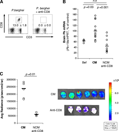
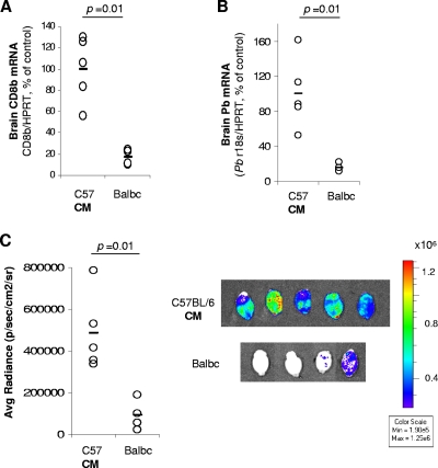
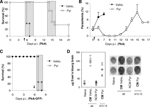
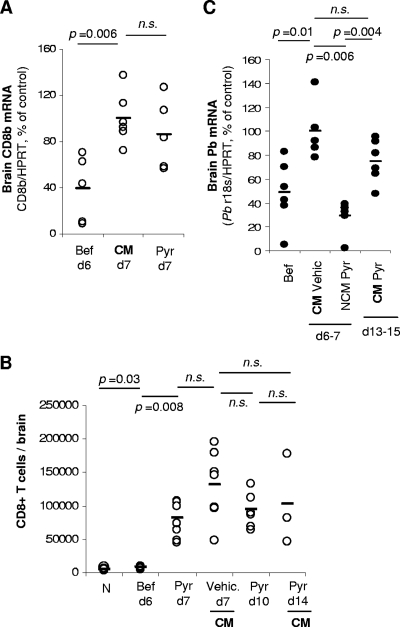
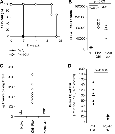
Cerebral malaria is the most severe complication of human infection with Plasmodium falciparum. It was shown that Plasmodium berghei ANKA-induced cerebral malaria was prevented in 100% of mice depleted of CD8+ T cells 1 day prior to the development of neurological signs. However, the importance of parasites in the brains of these mice was never clearly investigated. Moreover, the relevance of this model to human cerebral malaria has been questioned many times, especially concerning the relative importance of leukocytes versus parasitized erythrocytes sequestered in the brain. Here, we show that mice protected from cerebral malaria by CD8+ T-cell depletion have significantly fewer parasites in the brain. Treatment of infected mice with an antimalarial drug 15 to 20 h prior to the estimated time of death also protected mice from cerebral malaria without altering the number of CD8+ T cells in the brain. These mice subsequently developed cerebral malaria with parasitized red blood cells in the brain. Our results clearly demonstrated that sequestration of CD8+ T cells in the brain is not sufficient for the development of cerebral malaria in C57BL/6 mice but that the concomitant presence of parasitized red blood cells is crucial for the onset of pathology. Importantly, these results also demonstrated that the experimental cerebral malaria model shares many features with human pathology and might be a relevant model to study its pathogenesis.


Figures

FIG. 1.
FIG. 1.
Accumulation of parasites is reduced in the brains of CD8+ cell-depleted mice. Mice were infected with P. berghei ANKA (A and B) or a P. berghei ANKA line expressing luciferase (C). Groups of mice were treated with anti-CD8 antibodies 20 to 24 h before CM onset. (A) Depletion was checked by flow cytometry of blood lymphocytes 5 h after antibody injection. Data represent means ± SD (n = 5 mice per group). (B) When control mice developed CM, brains were harvested after intracardiac perfusion, and parasite mRNA expression was quantified by qRT-PCR. A group of mice was also analyzed before being treated with anti-CD8 antibodies (Bef). Each symbol represents one individual mouse from a pool of two independent experiments. (C) Mice were infected with luciferase-expressing P. berghei ANKA. When control mice developed CM, brain-accumulated pRBC were visualized after luciferin injection followed by intracardiac perfusion. n.s., not significantly different (Mann-Whitney test).
FIG. 2.
FIG. 2.
Accumulation of parasites is reduced in the brains of BALB/c mice. C57BL/6 (C57) or BALB/c mice were infected with P. berghei ANKA (A and B) or luciferase-expressing P. berghei ANKA (C). C57BL/6 mice were sacrificed when showing signs of CM (day 6 p.i.), and BALB/c mice were sacrificed on day 7 p.i., and brains were harvested after intracardiac perfusion. CD8β mRNA expression (A) and parasite mRNA expression (B) were quantified by qRT-PCR. (C) Brain-accumulated pRBC were visualized after luciferin injection followed by intracardiac perfusion. Each symbol represents one individual mouse. Data are representative of two independent experiments.
FIG. 3.
FIG. 3.
P. berghei ANKA-infected mice treated with antimalarial drugs do not develop ECM in the following days. P. berghei ANKA (A, B, and D)- or P. berghei ANKA-GFP (C)-infected mice were treated with pyrimethamine (Pyr) or vehicle (Vehic.) 15 to 20 h before the onset of CM (arrow): at the end of day 5 p.i. for P. berghei ANKA- and at day 6 p.i. for P. berghei ANKA-GFP-infected mice. The effects of vehicle or pyrimethamine treatment on survival (A and C) and parasitemia (B) are shown. Shaded areas indicate the times when mice displayed and died with CM symptoms. Data are representative of two independent experiments (n = 5 mice per group). (D) Qualitative (right) and quantitative (left) brain capillary permeability was assessed after intracardiac perfusion of Evans blue-injected mice. Naive (N), CM (vehicle-treated), and NCM (Pyr-treated) mice at day 6 p.i. and CM (Pyr-treated) mice at days 13 to 15 of infection were analyzed. Data represent the pooled results of two independent experiments. Each symbol represents one individual mouse.
FIG. 4.
FIG. 4.
Mice protected from ECM still accumulate CD8+ T cells in the brain but not pRBC. P. berghei ANKA-infected mice were treated with pyrimethamine (Pyr) or vehicle (Vehic.) 20 h before the onset of CM. Brains were harvested after intracardiac perfusion at different days p.i., before (Bef) or after pyrimethamine treatment (A to C). Sequestration of CD8+ T cells was assessed either by qRT-PCR (A) or by flow cytometry (B) in independent experiments. (C) Brain sequestration of parasites was assessed by qRT-PCR. Data are representative of three independent experiments. Each symbol represents one individual mouse. n.s., not significantly different (Mann-Whitney test).
FIG. 5.
FIG. 5.
CD8+ T cells but not P. berghei NK65 parasites accumulate in the brain in a non-CM model. Mice were infected i.p. with P. berghei ANKA or P. berghei NK65. (A) Survival of P. berghei NK65-infected mice is shown. (B to D) At day 7 p.i., when P. berghei ANKA-infected mice developed CM, brains were harvested after intracardiac perfusion. In independent experiments, sequestration of CD8+ T cells was assessed by flow cytometry (B), quantitative brain capillary permeability was assessed in Evans blue-injected mice (C), and parasite mRNA expression was quantified by qRT-PCR (D). Data are representative of two independent experiments. Each symbol represents one individual mouse. n.s., not significantly different (Mann-Whitney test).

References

    1. Adams, S., H. Brown, and G. Turner. 2002. Breaking down the blood-brain barrier: signaling a path to cerebral malaria? Trends Parasitol. 8:360-366. - PubMed
    1. Amante, F. H., A. C. Stanley, L. M. Randall, Y. Zhou, A. Haque, K. McSweeney, A. P. Waters, C. J. Janse, M. F. Good, G. R. Hill, and C. R. Engwerda. 2007. A role for natural regulatory T cells in the pathogenesis of experimental cerebral malaria. Am. J. Pathol. 171:548-559. - PMC - PubMed
    1. Bagot, S., F. Nogueira, A. Collette, V. do Rosário, F. Lemonier, P. A. Cazenave, and S. Pied. 2004. Comparative study of brain CD8+ T cells induced by sporozoites and those induced by blood-stage Plasmodium berghei ANKA involved in the development of cerebral malaria. Infect. Immun. 72:2817-2826. - PMC - PubMed
    1. Belnoue, E., M. Kayibanda, A. M. Vigário, J. C. Deschemin, N. van Rooijen, M. Viguier, G. Snounou, and L. Rénia. 2002. On the pathogenic role of brain-sequestered αβCD8+ T cells in experimental cerebral malaria. J. Immunol. 169:6369-6375. - PubMed
    1. Berendt, A. R., G. D. Tumer, and C. I. Newbold. 1994. Cerebral malaria: the sequestration hypothesis. Parasitol. Today 10:412-414. - PubMed

Publication types