. 2010 Sep;78(9):3871-82.
doi: 10.1128/IAI.00204-10. Epub 2010 Jul 6.

Conidia but not yeast cells of the fungal pathogen Histoplasma capsulatum trigger a type I interferon innate immune response in murine macrophages

Affiliations

Conidia but not yeast cells of the fungal pathogen Histoplasma capsulatum trigger a type I interferon innate immune response in murine macrophages

Diane O Inglis et al. Infect Immun. 2010 Sep.

Abstract

Histoplasma capsulatum is the most common cause of fungal respiratory infections and can lead to progressive disseminated infections, particularly in immunocompromised patients. Infection occurs upon inhalation of the aerosolized spores, known as conidia. Once inside the host, conidia are phagocytosed by alveolar macrophages. The conidia subsequently germinate and produce a budding yeast-like form that colonizes host macrophages and can disseminate throughout host organs and tissues. Even though conidia are the predominant infectious particle for H. capsulatum and are the first cell type encountered by the host during infection, very little is known at a molecular level about conidia or about their interaction with cells of the host immune system. We examined the interaction between conidia and host cells in a murine bone-marrow-derived macrophage model of infection. We used whole-genome expression profiling and quantitative reverse transcription-PCR (qRT-PCR) to monitor the macrophage signaling pathways that are modulated during infection with conidia. Our analysis revealed that type I interferon (IFN)-responsive genes and the beta type I IFN (IFN-beta) were induced in macrophages during infection with H. capsulatum conidia but not H. capsulatum yeast cells. Further analysis revealed that the type I IFN signature induced in macrophages in response to conidia is independent of Toll-like receptor (TLR) signaling and the cytosolic RNA sensor MAVS but is dependent on the transcription factor interferon regulatory factor 3 (IRF3). Interestingly, H. capsulatum growth was restricted in mice lacking the type I IFN receptor, indicating that an intact host type I IFN response is required for full virulence of H. capsulatum in mice.


Figures

FIG. 1.
FIG. 1.
Conidia are internalized by macrophages and germinate intracellularly to give rise to yeast cells. (A) Differential interference contrast (DIC) image (left) and immunofluorescence staining (right) of macrophages infected with conidia of the G217B strain. Both internal and external conidia are stained in blue with calcofluor, whereas only external conidia are stained in red with anti-Histoplasma antibodies. Macrophages are stained green using concanavalin A-FITC. (B) Periodic acid-Schiff (PAS) staining of conidia that have germinated and are producing yeast cells within macrophages 24 hpi. With PAS, conidia typically stain a darker magenta color than yeast cells. Three representative yeast cells are indicated with arrowheads, and two representative conidia are indicated with arrows.
FIG. 2.
FIG. 2.
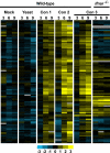
Heat map of type I IFN response genes induced by macrophages infected with Histoplasma conidia but not yeast. C57BL/6 (WT) macrophages were subjected to either mock infection, infection with H. capsulatum yeast cells, or infection with two independent preparations of H. capsulatum conidia (Con 1 and Con 2). WT and ifnar1/ macrophages were mock infected (data not shown) or infected with a third preparation of G217B conidia (Con 3). At 3, 6, and 9 hpi, macrophages were subjected to gene expression profiling. Genes with statistically significant induction in two independent wild-type macrophage infection experiments relative to the mock infection are shown. Yellow indicates gene upregulation, blue indicates downregulation, and black indicates no change. The color bar indicates the log2 scale.
FIG. 3.
FIG. 3.
Expression level of IFN-β in conidium- and yeast-infected macrophages. (A) qRT-PCR analysis to determine fold induction of IFN-β was performed on macrophage samples at various time points after infection with G217B conidia at an MOI of 10. (B) qRT-PCR analysis to determine fold induction of IFN-β at 3 hpi in macrophages infected with live or heat-killed (HK) conidia or with live yeast cells at an MOI of 10. (C and D) Macrophage lysates were subjected to qRT-PCR to detect relative induction of IFN-β after mock infection or infection with G217B, G184AR, G184AS, G186AR, or G186AS conidia (C) or yeast cells (D) at an MOI of 10. ND, not determined.
FIG. 4.
FIG. 4.
The type I IFN response to conidia is dependent on IRF3 and independent of MyD88, TRIF, and MAVS. qRT-PCR was used to determine fold IFN-β induction in irf3/ macrophages (A), myd88/ trif/ macrophages (B), and mavs+/ and mavs/ macrophages (C) infected with G217B conidia at an MOI of 10.
FIG. 5.
FIG. 5.
Induction of Ifi205 in AvMs infected with G217B conidia. qRT-PCR analysis to determine fold induction of Ifi205 was performed on AvM samples 4 h after infection with G217B conidia or yeast cells at an MOI of 10. Fold changes are calculated relative to the level in the mock-infected control. Error bars represent the standard error of the mean for replicate qRT-PCRs. The data shown include two (yeast cell A and B) or three (conidia A, B, and C) biological replicates.
FIG. 6.
FIG. 6.
Host type I IFN signaling is required for maximal fungal burden in host tissues following infection with H. capsulatum. WT and ifnar1/ mice were subjected to intranasal infection with G217B conidia or yeast cells. Two representative conidial infections and one representative yeast cell infection are shown. Lungs from infected animals were harvested at the indicated hours (h) or days (d) postinfection and assessed for CFU. Horizontal bars indicate mean log10 CFU values. Significant P values (P ≤ 0.05) for WT versus ifnar1/ comparisons are indicated on the figure.
FIG. 7.
FIG. 7.
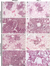
Wild-type mice have a more extensive inflammatory response to H. capsulatum conidia than ifnar1/ mice. Shown are hematoxylin-and-eosin-stained lung sections from mice infected with G217B conidia. Panels A and B are low-power images of representative inflammatory foci at 5 dpi in WT (A) or ifnar/ (B) mice. WT inflammatory foci are larger and more densely packed with immune cells. Scale bar, 200 μm. Panels C and D are high-power views of boxed regions from panels A and B. WT infiltrate contains many neutrophils, macrophages and eosinophils, with giant cells (GC) also present. Scale bar, 20 μm. (E and F) Low-power images of representative inflammatory foci at 14 dpi in WT (E) or ifnar/ (F) mice. Again, WT mouse inflammatory foci are larger and more densely packed than those of ifnar/ mice. Scale bar, 200 μm. Panels G and H are high-power views of boxed regions from panels E and F. The WT shows more densely organized macrophage and lymphocytic inflammation. Scale bar, 20 μm.
FIG. 8.
FIG. 8.
Giant cell formation in conidium-infected lungs of wild-type mice. Shown is high magnification of a giant cell containing H. capsulatum macroconidia (black arrows), microconidia (white arrowheads), and yeast cells (black arrowheads) observed in the lungs of conidium-infected WT mouse strains at 5 dpi. Conidial forms could represent ungerminated cells or conidial remnants that persist after germination. Scale bar, 20 μm.

References

    1. Aderem, A. 2003. Phagocytosis and the inflammatory response. J. Infect. Dis. 187(Suppl. 2):S340-S345. - PubMed
    1. Akira, S., S. Uematsu, and O. Takeuchi. 2006. Pathogen recognition and innate immunity. Cell 124:783-801. - PubMed
    1. Anderson, K. L., and S. Marcus. 1968. Sporulation characteristics of Histoplasma capsulatum. Mycopathol. Mycol. Appl. 36:179-187. - PubMed
    1. Andreas, S., S. Heindl, C. Wattky, K. Moller, and R. Ruchel. 2000. Diagnosis of pulmonary aspergillosis using optical brighteners. Eur. Respir. J. 15:407-411. - PubMed
    1. Auerbuch, V., D. G. Brockstedt, N. Meyer-Morse, M. O'Riordan, and D. A. Portnoy. 2004. Mice lacking the type I interferon receptor are resistant to Listeria monocytogenes. J. Exp. Med. 200:527-533. - PMC - PubMed

Publication types

Associated data