. 2010 Sep;78(9):3753-62.
doi: 10.1128/IAI.00502-09. Epub 2010 Jul 6.

The lung is an important site for priming CD4 T-cell-mediated protective immunity against gastrointestinal helminth parasites

Affiliations

The lung is an important site for priming CD4 T-cell-mediated protective immunity against gastrointestinal helminth parasites

Marina Harvie et al. Infect Immun. 2010 Sep.

Abstract

The rodent hookworm Nippostrongylus brasiliensis typically infects its host by penetrating the skin and rapidly migrating to the lungs and gut. Following primary infection, immunocompetent mice become highly protected from reinfection with N. brasiliensis, with the numbers of worms gaining access to the lungs and gut being reduced by up to 90%. We used green fluorescent protein/interleukin-4 (IL-4) reporter mice and truncated infection studies to identify both the tissue site and mechanism(s) by which the host protects itself from reinfection with N. brasiliensis. Strikingly, we demonstrated that the lung is an important site for priming immune protection. Furthermore, a lung-initiated, CD4 T-cell-dependent, and IL-4- and STAT6-dependent response was sufficient to confer protection against reinfection. In conclusion, vaccination strategies which seek to break the cycle of reinfection and egg production by helminths such as hookworms can include strategies which directly stimulate Th2 responses in the lung.


Figures

FIG. 1.
FIG. 1.
Reduction in worm burden correlates with induction of CD4 GFP+ Th2 immune response in mice reinfected with N. brasiliensis. (A) Number of larvae migrating from site of skin infection. G4/IL-4 mice (n = 3 per time point) were infected i.d. in the ear pinna with 600 L3 N. brasiliensis worms (open squares) or left uninfected (closed squares), rested, and challenged by i.d. reinfection with 600 L3 N. brasiliensis worms. At the time points indicated, viable larvae that migrated out of excised ears were enumerated. Values are expressed as percent burden ± the standard error of the mean (SEM) and are representative of three experiments. (B) Representative photomicrograph of a mouse ear 5 min after the injection of CFSE-labeled L3 N. brasiliensis worms. Inset, differential interference contrast versus fluorescence microscopy of the same worms. The worms pictured are 0.5 mm in length. (C) Parasite burden during s.c. primary N. brasiliensis infection. G4/IL-4 mice (n = 4) were primed with 600 L3 N. brasiliensis worms s.c. Lung (closed squares) and gut (open squares) tissues were harvested, and viable worms that migrated out were enumerated. The values shown represent the mean number of worms recovered ± the SEM and are representative of three independent experiments. (D) Parasite burden during N. brasiliensis reinfection. G4/IL-4 (n = 4) mice were primed with 600 L3 N. brasiliensis worms s.c., rested, and reinfected s.c. with 600 L3 N. brasiliensis worms. Lung (closed squares) and gut (open squares) tissues were harvested, and viable worms that migrated out were enumerated. The values shown represent the mean number of worms recovered ± the SEM and are representative of three independent experiments. (E) GFP+ CD4 T-cell responses during primary s.c. N. brasiliensis infection. Mediastinal (closed squares) and mesenteric (open squares) lymph nodes from s.c. N. brasiliensis-infected animals (n = 4) were harvested and surface stained for CD3 and CD4. The values shown represent the mean number of cells ± the SEM and are representative of at least three independent experiments. (F) GFP+ CD4 T-cell responses during N. brasiliensis reinfection. Mediastinal (closed squares) and mesenteric (open squares) lymph nodes from s.c. N. brasiliensis-infected animals (n = 4) were harvested and surface stained for CD3 and CD4. The values shown represent the mean number of cells ± the SEM and are representative of at least three independent experiments.
FIG. 2.
FIG. 2.
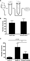
The skin is not the site of protection from reinfection. (A) Schematic representation of in vitro skin migration assay. Skin harvested from naïve or previously s.c. infected mice (n = 4 per group) was suspended over PBS. iL3 worms were placed on the skin samples, and those that migrated through were enumerated. (B) L3 worm skin migration efficiency in vitro. L3 N. brasiliensis worms able to migrate through naïve or immune skin (as described for panel A) were enumerated. The values shown represent the mean number of larvae that migrated ± the SEM. (C) Priming with dead worms compromises protection. G4/IL-4 mice (n = 3 per group) were inoculated i.d. in the ear pinna with 600 dead or live L3 N. brasiliensis worms, rested, and challenged s.c. with 600 live L3 N. brasiliensis worms. Lungs were excised on day 2 postinfection, and the viable worms present were enumerated using the migration assay described in Materials and Methods. Data show the mean number of worms that migrated out ± the SEM. All experiments are representative of three experiments.
FIG. 3.
FIG. 3.
Schematic representation of the truncated infection strategy used for selective infection of lung and gut tissues. (A) Skin and lung priming. Mice were infected with ∼600 L3 N. brasiliensis worms s.c. and given an anthelminthic daily by gavage from day 2 to day 9 to prevent the establishment of L4/L5 worms in the gut and prevent the induction of a gut-localized immune response to N. brasiliensis. Mice were s.c. reinfected with 600 L3 N. brasiliensis worms at day 30 postinfection. (B) Lung priming. Mice were i.n. administered ∼200 L4 N. brasiliensis worms and given an anthelminthic daily by gavage from day 2 to day 9 to prevent the establishment of L4/L5 worms in the gut and prevent the induction of a gut-localized immune response to N. brasiliensis. Mice were s.c. reinfected with 600 L3 N. brasiliensis worms on day 30 postinfection. (C) Gut priming. Mice were infected with ∼300 L5 N. brasiliensis worms by gavage. Mice were s.c. challenged with 600 L3 N. brasiliensis worms on day 30 postinfection. (D) Skin, lung, and gut priming. Mice were infected with ∼600 L3 N. brasiliensis worms s.c. Mice were s.c. reinfected with 600 L3 N. brasiliensis worms on day 30 postinfection. (E) No-priming (naive control) naïve mice were s.c. infected with ∼600 L3 N. brasiliensis worms at the same time as the groups shown in panels A, B, C, and D were s.c. reinfected to enable comparison of worm burdens and calculation of the level of protection.
FIG. 4.
FIG. 4.
Effect of truncated skin, lung, and gut primary N. brasiliensis infection on the development of immunity to reinfection. G4/IL-4 mice (n = 4) were infected via the skin and lungs, the lungs only, the gut only, or whole infection. The anthelminthic was given from day 2 to day 9 to the skin-and-lung infection and lung infection only groups. Following the 30-day rest period postinfection, mice were s.c. infected with 600 N. brasiliensis L3 worms and on day 2 their lungs were excised and the worms that migrated out were enumerated. The values shown represent the mean number of larvae present in the lung ± the SEM. The data shown are representative of three separate experiments. (B) Worm burdens in the gut following priming with distinct phases of the N. brasiliensis life cycle. G4/IL-4 mice (n = 4) were infected via the skin and lungs, the lungs only, the gut only, or whole infection, and an anthelminthic was given from day 2 to day 9 to the skin-and-lung and lung-only groups. Following a rest period, mice were s.c. infected with 600 N. brasiliensis L3 worms and on day 6, their guts were excised and the worms that migrated out were enumerated. The values shown represent the mean number of larvae present in the gut ± the SEM. The data shown are representative of three separate experiments. (C) GFP+ CD4 T-cell responses in mediastinal lymph nodes during distinct phases of the N. brasiliensis life cycle. Mediastinal lymph nodes from animals (n = 4) infected via the skin and lungs, the lungs only, the gut only, or whole infection were harvested on day 2 and surface stained for CD3 and CD4. The values shown represent mean cell numbers ± the SEM and are representative of three independent experiments. (D) GFP+ CD4 T-cell responses in mesenteric lymph nodes during distinct phases of the N. brasiliensis life cycle. Mesenteric lymph nodes from animals (n = 4/group) infected via the skin and lungs, the lungs only, the gut only, or whole infection were harvested on day 6 and surface stained for CD3 and CD4. The values shown represent mean cell numbers ± the SEM and are representative of three independent experiments.
FIG. 5.
FIG. 5.
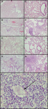
Histological analysis of the lungs of N. brasiliensis-infected mice. BALB/c mice (n = 3/group) were infected s.c. with 600 N. brasiliensis worms and rested for 30 days before being reinfected s.c. with 600 iL3 N. brasiliensis worms. Lung tissues were harvested and fixed at the indicated time points before being separated into 4-μm sections and stained with H&E to visualize inflammatory cells. (A and E) Lung tissue from naïve mice at ×40 and ×200 magnifications, respectively. (B and F) Lung tissue from a primary s.c. N. brasiliensis-infected animal at day 9 postinfection at ×40 and ×200 magnifications, respectively. (C and G) Lung tissue from an N. brasiliensis-infected animal at day 29 after s.c. infection (prior to reinfection) at ×40 and ×200 magnifications, respectively. (D, H, and I) Lung tissue taken from mice s.c. reinfected with N. brasiliensis at day 6 after s.c. reinfection at ×40, ×200, and ×1,000 magnifications, respectively. The arrow in panel I indicates an N. brasiliensis worm.
FIG. 6.
FIG. 6.
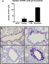
Mucus production in the airways of N. brasiliensis-infected mice. BALB/c mice (n = 3/group) were infected s.c. with 600 worms and rested for 30 days before being reinfected s.c. with 600 iL3 N. brasiliensis worms. Lung tissues were harvested and fixed at the indicated time points before being separated into 4-μm sections and stained with PAS and alcian blue to visualize mucus-secreting goblet cells. (A) The number of mucus producing cells per bronchiole was determined by counting the PAS-positive cells in all bronchioles 200 to 600 μm in size per lung section. Data represent the mean number of mucus-producing cells per bronchiole ± the SEM. (B) Representative bronchiole from a naive animal, ×400 magnification. (C) Representative bronchiole from an s.c. N. brasiliensis-infected animal at day 9 postinfection, ×400 magnification. (D) Representative bronchiole from an N. brasiliensis-infected animal at day 29 after s.c. infection (prior to reinfection [PTR]), ×400 magnification. (E) Representative bronchiole from an s.c. N. brasiliensis-reinfected animal at day 6 after reinfection, ×400 magnification.

References

    1. Agrawal, S., A. Agrawal, B. Doughty, A. Gerwitz, J. Blenis, T. Van Dyke, and B. Pulendran. 2003. Cutting edge: different Toll-like receptor agonists instruct dendritic cells to induce distinct Th responses via differential modulation of extracellular signal-regulated kinase-mitogen-activated protein kinase and c-Fos. J. Immunol. 171:4984-4989. - PubMed
    1. Aitken, R., P. S. Coulson, and R. A. Wilson. 1988. Pulmonary leukocytic responses are linked to the acquired immunity of mice vaccinated with irradiated cercariae of Schistosoma mansoni. J. Immunol. 140:3573-3579. - PubMed
    1. Brown, A., N. Girod, E. E. Billett, and D. I. Pritchard. 1999. Necator americanus (human hookworm) aspartyl proteinases and digestion of skin macromolecules during skin penetration. Am. J. Trop. Med. Hyg. 60:840-847. - PubMed
    1. Camberis, M., G. Le Gros, and J. Urban, Jr. 2003. Animal model of Nippostrongylus brasiliensis and Heligmosomoides polygyrus. Curr. Protoc. Immunol. Chapter 19:Unit 19.12. - PubMed
    1. Coulson, P. S., and R. A. Wilson. 1997. Recruitment of lymphocytes to the lung through vaccination enhances the immunity of mice exposed to irradiated schistosomes. Infect. Immun. 65:42-48. - PMC - PubMed

Publication types

LinkOut - more resources