. 2010 Sep;30(17):4245-53.
doi: 10.1128/MCB.00218-10. Epub 2010 Jul 6.

Deficiency in a glutamine-specific methyltransferase for release factor causes mouse embryonic lethality

Affiliations

Deficiency in a glutamine-specific methyltransferase for release factor causes mouse embryonic lethality

Peng Liu et al. Mol Cell Biol. 2010 Sep.

Abstract

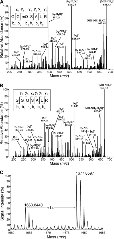
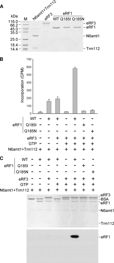
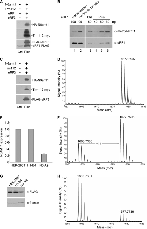
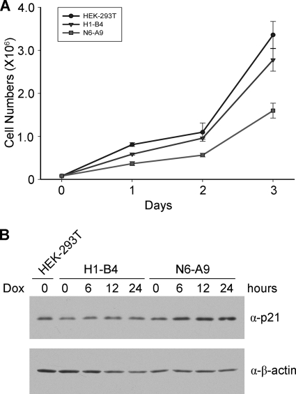
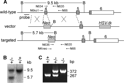
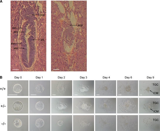
Biological methylation is a fundamental enzymatic reaction for a variety of substrates in multiple cellular processes. Mammalian N6amt1 was thought to be a homologue of bacterial N(6)-adenine DNA methyltransferases, but its substrate specificity and physiological importance remain elusive. Here, we demonstrate that N6amt1 functions as a protein methyltransferase for the translation termination factor eRF1 in mammalian cells both in vitro and in vivo. Mass spectrometry analysis indicated that about 70% of the endogenous eRF1 is methylated at the glutamine residue of the conserved GGQ motif. To address the physiological significance of eRF1 methylation, we disrupted the N6amt1 gene in the mouse. Loss of N6amt1 led to early embryonic lethality. The postimplantation development of mutant embryos was impaired, resulting in degeneration around embryonic day 6.5. This is in contrast to what occurs in Escherichia coli and Saccharomyces cerevisiae, which can survive without the N6amt1 homologues. Thus, N6amt1 is the first glutamine-specific protein methyltransferase characterized in vivo in mammals and methylation of eRF1 by N6amt1 might be essential for the viability of early embryos.


Figures

FIG. 1.
FIG. 1.
Identification of Gln185 methylation of eRF1 in vivo in mammalian cells. (A) ESI-MS/MS spectrum of methylated 702.34-Da fragments [heptapeptide GG(mQ)SALR, amino acids 183 to 189] from endogenous eRF1. eRF1 was purified by immunoprecipitation from the HEK-293T cell extract using anti-eRF1 polyclonal antibodies, separated by PAGE, and subjected to in-gel digestion with trypsin prior to ESI-MS/MS. The fragmentation pattern established the heptapeptide sequence indicated. (B) ESI-MS/MS spectrum of methylation-free 688.32-Da fragments from recombinant eRF1 purified from E. coli. (C) MALDI-TOF MS spectrum of endogenous eRF1 of HEK-293T cells. eRF1 was affinity purified with specific antibodies. The peak at m/z 1,663.84 is from the unmethylated peptide (sequence TVDLPKKHGRGGQSAL, amino acids 173 to 188) generated from chymotrypsin digestion. The peak at m/z 1,677.86 represents the methylated peptide.
FIG. 2.
FIG. 2.
Characterization of the in vitro methylation activity of recombinant N6amt1. (A) Recombinant proteins used for the methylation assay. 6×His-tagged N6amt1 coexpressed FLAG-Trm112, GST-eRF3, and wild-type (WT) and mutant 6×His-eRF1 proteins were purified from E. coli and examined by Coomassie staining. (B) Methyltransferase activity detected by scintillation counting. eRF1 substrates were incubated with N6amt1 in the presence or absence of other factors using [methyl-3H]AdoMet as the methyl donor, and the proteins were then precipitated by trichloroacetic acid (TCA) for the measurement of methyl-3H incorporation. Averages from three independent experiments ± standard deviations are shown. (C) Methyl-transferase activity detected by fluorography. After the methylation reaction in the presence of radioactive [methyl-3H]AdoMet, the proteins were separated by SDS-PAGE and visualized by Coomassie staining (upper panel), and the same gel was subjected to fluorography (lower panel). Note that only the wild-type eRF1 was radioactively labeled.
FIG. 3.
FIG. 3.
Characterization of the in vivo methylation activity of N6amt1 in transfected HEK-293T cells. (A) Detection of transfected proteins by Western analysis. HEK-293T cells were transiently transfected with the eRF1-FLAG expression construct alone (“Ctrl” sample) and with the constructs for HA-N6amt1, Trm112-myc, and FLAG-eRF3 (“Plus” sample). Epitope-specific antibodies were used in the detection. (B) Increased methylation of eRF1 when cotransfected with HA-N6amt1, Trm112-myc, and FLAG-eRF3. The ectopic eRF1 was immunoprecipitated from the cell extract using anti-FLAG M2 beads prior to Western analysis. Bacterial recombinant eRF1 protein samples, unmethylated and methylated in vitro, were loaded as a control to monitor preferential detection of the methylated form by the anti-methyl-eRF1 (α-methyl-eRF1) antibody (lanes 1 and 2). Different amounts (40, 50, and 60 ng) of the “Plus” eRF1 sample were loaded for quantification of the methylated eRF1. The antibodies used are indicated to the right. α-eRF1, anti-eRF1 antibody. (C) Western detection of N6amt1 and its cofactors transfected into HEK-293T cells for the methylation analysis on endogenous eRF1. Epitope-specific antibodies were used. (D) MALDI-TOF MS analysis for Gln185 methylation of endogenous eRF1 from HEK-293T cells transfected with N6amt1 and its cofactors (“Plus” sample). The endogenous eRF1 protein was immunopurified with anti-eRF1 polyclonal antibodies from transfected HEK-293T cells, separated, and in-gel digested with chymotrypsin prior to MALDI-TOF MS analysis. The peak at m/z 1,677.69 corresponds to methylated peptides containing Gln185 (Fig. 1C). The peak for unmethylated peptide was not detected. (E) N6AMT1 depletion in knockdown cell line N6-A9 verified by real-time PCR. siRNA expression was induced by addition of doxycycline. H1-B4 is a control cell line harboring an empty siRNA vector. (F) N6AMT1 depletion in knockdown cell line N6-A9 verified by Western blotting. (G) MALDI-TOF MS analysis for Gln185 methylation of FLAG-eRF1 in H1-B4 cells. α-FLAG, anti-FLAG antibody; α-β-actin, anti-β-actin antibody. (H) MALDI-TOF MS analysis for Gln185 methylation of FLAG-eRF1 in N6-A9 cells.
FIG. 4.
FIG. 4.
Reduced proliferation of N6AMT1 knockdown cells and modest upregulation of p21. (A) Growth curve of knockdown (N6-A9) and control (HEK-293T and H1-B4) cells in medium with doxycycline. (B) Expression of p21 upon doxycycline (Dox) induction in N6-A9 cells. Western blotting was carried out three times, and representative results are shown.
FIG. 5.
FIG. 5.
Targeted disruption of the N6amt1 gene in mice. (A) Targeting strategy to generate the mutant N6amt1 allele. Numbered gray boxes represent coding exons, and open boxes represent the 3′ untranscribed region (UTR). B, BamHI restriction site; Neo, neomycin resistance gene; HSV-tk, herpes simplex virus thymidine kinase gene. The locations of the Southern probe and the expected BamHI fragments are indicated. PCR primers used for genotyping are indicated with arrows. (B) Genotyping of ES clones by Southern blot analysis. Genomic DNA was digested with BamHI. The 9.5-kb and 5.7-kb bands were derived from the wild-type and targeted alleles, respectively. (C) Genotyping of mouse embryos by PCR. Heterozygous mice were intercrossed, and E3.5 blastocysts were collected. The 267-bp and 372-bp PCR products were derived from the wild-type and targeted alleles, respectively.
FIG. 6.
FIG. 6.
Early embryonic lethality caused by deficiency in N6amt1. (A) H&E staining of longitudinal sections of E6.5 embryos from intercrosses of N6amt1+/ mice. Typical embryos with normal (left) and abnormal (right) morphologies are shown. The normal E6.5 embryo has initiated the formation of a primitive streak (ps) and developed an elongated egg cylinder. The abnormal embryo is much smaller in overall size. The ectoplacental cone (epc) is underdeveloped, and no primitive streak forms. Endoderm and ectoderm cannot be distinguished. xen, extraembryonic endoderm; xec, extraembryonic ectoderm; een, embryonic endoderm; eec, embryonic ectoderm. Scale bar, 50 μm. (B) Outgrowth of blastocysts from N6amt1+/ intercrosses in vitro. E3.5 blastocysts were collected and cultured in ES medium for 9 days. Genotypes were determined by genomic PCR. ICM and trophoblastic giant cells (TGC) in outgrowth at day 9 are pointed out. Scale bars, 100 μm.

References

    1. Antonarakis, S. E., R. Lyle, S. Deutsch, and A. Reymond. 2002. Chromosome 21: a small land of fascinating disorders with unknown pathophysiology. Int. J. Dev. Biol. 46:89-96. - PubMed
    1. Bujnicki, J. M., and M. Radlinska. 1999. Is the HemK family of putative S-adenosylmethionine-dependent methyltransferases a “missing” zeta subfamily of adenine methyltransferases? A hypothesis. IUBMB Life 48:247-249. - PubMed
    1. Clarke, S. 1993. Protein methylation. Curr. Opin. Cell Biol. 5:977-983. - PubMed
    1. Dincbas-Renqvist, V., A. Engstrom, L. Mora, V. Heurgue-Hamard, R. Buckingham, and M. Ehrenberg. 2000. A post-translational modification in the GGQ motif of RF2 from Escherichia coli stimulates termination of translation. EMBO J. 19:6900-6907. - PMC - PubMed
    1. Figaro, S., N. Scrima, R. H. Buckingham, and V. Heurgue-Hamard. 2008. HemK2 protein, encoded on human chromosome 21, methylates translation termination factor eRF1. FEBS Lett. 582:2352-2356. - PubMed

Publication types

Substances