. 2010 Sep;30(17):4339-53.
doi: 10.1128/MCB.00545-10. Epub 2010 Jul 6.

The early isoform of disabled-1 functions independently of Reelin-mediated tyrosine phosphorylation in chick retina

Affiliations

The early isoform of disabled-1 functions independently of Reelin-mediated tyrosine phosphorylation in chick retina

Zhihua Gao et al. Mol Cell Biol. 2010 Sep.

Abstract

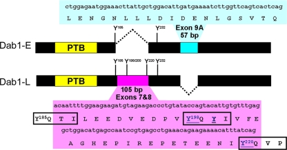
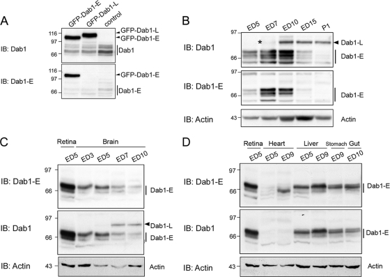
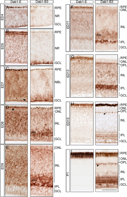
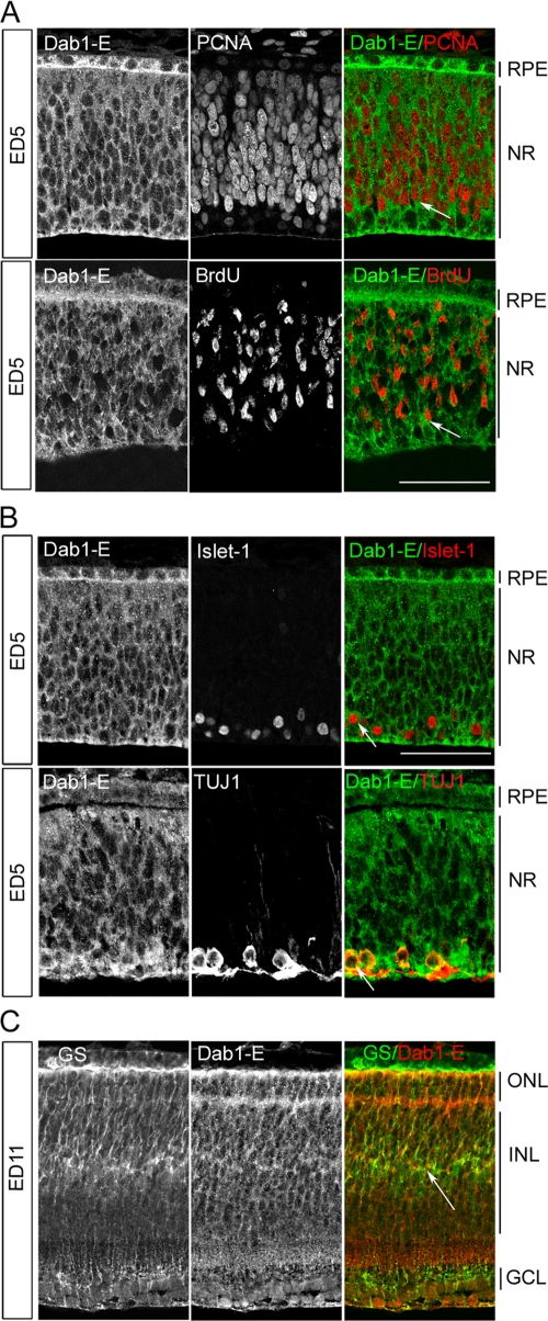
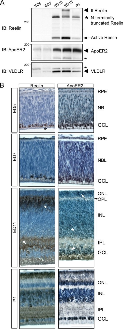
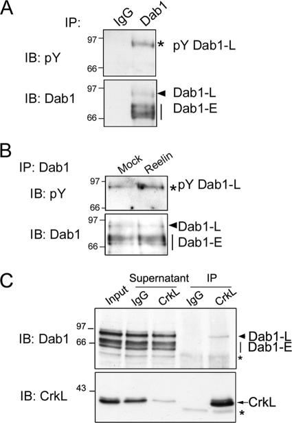
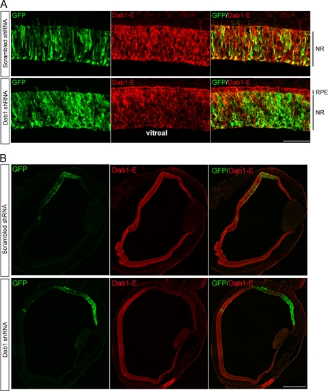
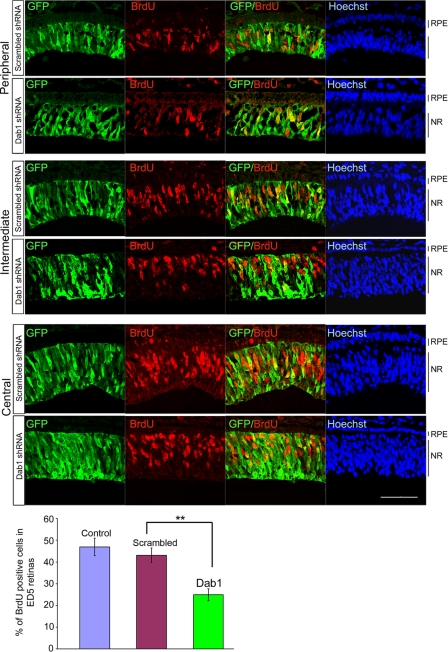
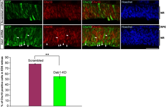
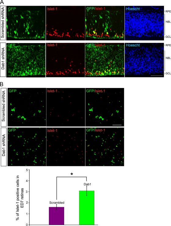
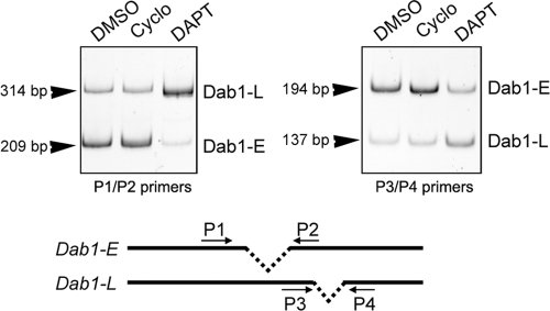
The Reelin-Disabled-1 (Dab1) signaling pathway plays a key role in the positioning of neurons during brain development. Two alternatively spliced Dab1 isoforms have been identified in chick retina and brain: Dab1-E, expressed at early stages of development, and Dab1-L (commonly referred to as Dab1), expressed at later developmental stages. The well-studied Dab1-L serves as an adaptor protein linking Reelin signal to its downstream effectors; however, nothing is known regarding the role of Dab1-E. Here we show that Dab1-E is primarily expressed in proliferating retinal progenitor cells whereas Dab1-L is found exclusively in differentiated neuronal cells. In contrast to Dab1-L, which is tyrosine phosphorylated upon Reelin stimulation, Dab1-E is not tyrosine phosphorylated and may function independently of Reelin. Knockdown of Dab1-E in chick retina results in a significant reduction in the number of proliferating cells and promotes ganglion cell differentiation. Our results demonstrate a role for Dab1-E in the maintenance of the retinal progenitor pool and determination of cell fate.


Figures

FIG. 1.
FIG. 1.
Schematic diagram of exon exclusion and inclusion in Dab1 isoforms. The two exons deleted in Dab1-E but included in Dab1-L are shown in magenta; the exon included in Dab1-E but excluded from Dab1-L is shown in blue. The phosphotyrosine binding (PTB) domain common to both Dab1 isoforms is shown in yellow. Two tyrosines, at 185 and 232, are indicated in Dab1-E. Five tyrosines, at 185, 198, 200, 220, and 232, are indicated in Dab1-L. Alternative splicing converts Y185QTI (in Dab1-L) to Y185QVP (in Dab1-E). YQXI is a consensus Src family kinase phosphorylation site, whereas YXVP is a consensus Abl family kinase recognition site.
FIG. 2.
FIG. 2.
Dab1 expression in the chicken embryo. (A) Determination of anti-Dab1-E antibody specificity. Fifty μg cell lysates prepared from ED5 retinal cultures transfected with GFP (control), GFP-Dab1-E, or GFP-Dab1-L expression constructs were resolved by 8% SDS-PAGE and transferred to a polyvinylidene difluoride (PVDF) membrane. The membrane was immunostained with anti-Dab1 (raised against the C terminus of Dab1, top panel) or anti-Dab1-E (raised against the E-specific region, bottom panel) antibody. Anti-Dab1 antibody recognizes both Dab1-E and -L, whereas the anti-Dab1-E antibody specifically recognizes Dab1-E. The endogenous Dab1 and Dab1-E proteins are indicated by vertical bars. IB, immunoblot. (B) Western blot analysis of Dab1 in the chick retina. To maximize the separation between the Dab1 isoforms, 100 μg retinal cell lysates prepared from ED5, ED7, ED10, ED15, and P1 were resolved on a 20-cm by 20-cm 8% SDS acrylamide gel and transferred to a PVDF membrane. The membrane was immunostained with anti-Dab1 (top panel) or anti-Dab1-E (middle panel) antibody. The arrowhead indicates Dab1-L, whereas the vertical bars show the early form(s) of Dab1 (Dab1-E). The asterisk indicates a weakly stained Dab1-L band in ED7 retina. (C) Western blot analysis of Dab1 in the chick brain. Cell lysates prepared from ED5 chick retina, ED3 chick head, ED5, ED7, and ED10 chick brain were analyzed by Western blotting as indicated in panel A. (D) Western blot analysis of Dab1 in embryonic chick tissues. Cell lysates prepared from chick retina, heart, stomach, liver, and gut at different developmental stages (as indicated) were analyzed by Western blotting. Actin was used as a loading control.
FIG. 3.
FIG. 3.
Immunohistochemical analysis of Dab1 isoforms in the developing chick retina. Consecutive sections from formalin-fixed paraffin-embedded chick retina at different developmental stages (as indicated) were immunostained with anti-Dab1-E or anti-Dab1 B3 antibody. Sections were not counterstained in order to better visualize Dab1 staining. The arrowhead in panel C points to the reduced expression of Dab1-E in the GCL. In panel D, the asterisk indicates the reduced levels of Dab1-E in the emerging ONL. Abbreviations: RPE, retinal pigmented epithelium; NR, neural retina; NBL, neuroblastic layer; INL, inner nuclear layer; IPL, inner plexiform layer; GCL, ganglion cell layer; ONL, outer nuclear layer; OPL, outer plexiform layer; Am, amacrine. Scale bar, 50 μm.
FIG. 4.
FIG. 4.
Expression of Dab1-E in proliferating retinal progenitor cells and newly committed postmitotic cells. (A) Coimmunostaining of Dab1-E and PCNA or Dab1-E and BrdU in ED5 chick retina. Retinal sections were doubly stained with rabbit anti-Dab1-E (1:400) and either mouse anti-PCNA (1:5,000) or mouse anti-BrdU (1:50) antibodies, followed by donkey anti-rabbit and donkey anti-mouse secondary antibodies conjugated with fluorescent Alexa 488 and Alexa 555, respectively. Arrows indicate the colabeling of Dab1-E (cytoplasmic) and PCNA (nuclear) or BrdU (nuclear) in retinal cells. (B) Coimmunostaining of Dab1-E and Islet-1 or Dab1-E and TUJ1 in ED5 chick retina. Retinal sections were doubly stained with rabbit anti-Dab1-E and mouse anti-Islet-1 (1:1,500) or mouse anti-TUJ1 (1:2,000) antibodies as indicated in panel A. Arrows indicate the colabeling of Dab1-E and Islet-1 or TUJ1 in retinal cells. (C) Coimmunostaining analysis of Dab1-E and glutamine synthetase (GS) in ED11 chick retina. Retinal sections were doubly stained with rabbit anti-Dab1-E and mouse anti-GS (1:500) antibodies, followed by donkey anti-rabbit or donkey anti-mouse secondary antibodies conjugated with Alexa 555 and Alexa 488, respectively. Arrows indicate the colabeling of Dab1-E and GS in ED11 chick retina. Micrographs were collected with a Zeiss LSM 510 confocal microscope equipped with a 40× lens. For abbreviations, see the legend for Fig. 3. Scale bar, 50 μm.
FIG. 5.
FIG. 5.
Expression of Reelin, ApoER2, and VLDLR in the developing chick retina. (A) Western blot analysis of Reelin, ApoER2, and VLDLR in the developing chick retina. Western blotting of Reelin, ApoER2, and VLDLR was carried out as described in the legend for Fig. 2B. The membrane was immunostained with anti-Reelin (1:500, top panel), ApoER2 (1:2,000, middle panel), or VLDLR (1:200, bottom panel) antibodies, respectively. Actin was used as a loading control. The arrowhead in the top panel indicates full-length Reelin, whereas the large asterisk and the arrow indicate N-terminally truncated Reelin and active Reelin, respectively. Arrowheads in the middle and bottom panels indicate the most abundant forms of ApoER2 and VLDLR, whereas small asterisks indicate alternatively spliced isoforms. (B) Immunohistochemical analysis of Reelin and ApoER2 in the developing chick retina. Chick retinal sections at different developmental stages (as indicated) were immunostained with mouse anti-Reelin (1:500) or rabbit anti-ApoER2 (1:2,000) antibodies, and nuclei were labeled with hematoxylin. At ED5, the asterisk (left panel) indicates Reelin expression in ganglion cells. At ED11, the arrow and arrowhead (left panel) point to the expression of Reelin in bipolar and ganglion cells, respectively. The arrow, asterisk, and arrowhead (right panel) indicate ApoER2 expression in a subset of photoreceptors and amacrine and ganglion cells in ED11 chick retina. For abbreviations, see the legend for Fig. 3. Scale bar, 50 μm.
FIG. 6.
FIG. 6.
Examination of Dab1 tyrosine phosphorylation in the retina. (A) Tyrosine phosphorylation of Dab1-L in ED10 chick retina. Western blotting of anti-Dab1 antibody and IgG control immunoprecipitates from ED10 chick retinal extracts was carried out as for Fig. 2A. The membrane was immunostained with mouse anti-pY 4G10 and rabbit anti-Dab1 antibodies. (B) Reelin stimulates Dab1-L tyrosine phosphorylation but not that of Dab1-E in ED10 retinal cells. ED10 primary retinal cultures were treated with mock or Reelin-conditioned medium for 15 min, followed by cell lysis in RIPA buffer. Dab1 proteins were immunoprecipitated from the cell lysates and analyzed by Western blotting. Tyrosine-phosphorylated Dab1 was detected with a mouse anti-pY 4G10 antibody, and Dab1 identity was confirmed with an anti-Dab1 antibody. (C) Dab1-L but not Dab1-E associates with CrkL. Immunocomplexes were immunoprecipitated from ED10 chick retinal lysates by use of mouse anti-CrkL or mouse IgG control. The immunoprecipitates, supernatants, and 10% input were analyzed by Western blotting using anti-Dab1 and CrkL antibodies. Asterisks indicate nonspecific bands.
FIG. 7.
FIG. 7.
Knockdown of Dab1-E in the developing chick retina. (A) Knockdown of Dab-E in ED5 chick retina. Cryosections from ED5 chick retinas electroporated with RCAS-GFP-Scrambled or RCAS-GFP-Dab1 shRNA were coimmunostained with goat anti-GFP and rabbit anti-Dab1-E antibodies. (B) Knockdown of Dab1-E in ED7 chick retina. Cryosections from ED7 chick retinas electroporated with RCAS-GFP-scrambled or RCAS-GFP-Dab1 shRNA were doubly immunostained with goat anti-GFP and rabbit anti-Dab1-E antibodies. Micrographs were collected with a Zeiss LSM 510 confocal microscope equipped with a 40× (A) or 10× (B) lens. A 3 × 3 tile scan was applied to cover the entire retina in panel B. Abbreviations: RPE, retinal pigmented epithelium; NR, neural retina. Scale bar, 50 μm (A) or 500 μm (B).
FIG. 8.
FIG. 8.
BrdU incorporation in ED5 chick retinas electroporated with scrambled or Dab1 shRNA. BrdU and GFP were visualized by immunostaining with anti-mouse BrdU (1:100) and anti-goat GFP (1:2,000) antibodies. Sections were counterstained with Hoechst 33342 to label the nuclei. Representative images from peripheral, intermediate, and central zones from in ovo electroporated ED5 chick retinas are shown. Micrographs were collected with a Zeiss LSM 510 confocal microscope equipped with a 40× lens. Scale bar, 50 μm. The histogram shows the percentage of GFP+ cells that are positive for BrdU. Values were derived from 6 controls and 6 scrambled and 8 Dab1 knockdown embryos. No significant difference was found between the control and scrambled groups. There was a significant difference between the scrambled and Dab1-E knockdown groups. **, P < 0.01 (t test). Abbreviations: RPE, retinal pigmented epithelium; NR, neural retina.
FIG. 9.
FIG. 9.
Chx10 expression in ED5 chick retinas electroporated with scrambled or Dab1 shRNA. Chx10 and GFP were visualized by immunostaining with anti-sheep Chx10 (1:3,000) and anti-rabbit GFP (1:4,000) antibodies. Sections were counterstained with Hoechst 33342 to label the nuclei. Micrographs were collected from 4 eyes electroporated with scrambled shRNA and 4 eyes electroporated with Dab1 shRNA as described in the legend for Fig. 8. The arrows point to some of the GFP+ Chx10 cells observed upon electroporation with Dab1 shRNA. Scale bar, 50 μm. The histogram shows the percentage of GFP+ retinal cells that are positive for Chx10. There was a significant difference between the scrambled and Dab1-E knockdown groups. **, P < 0.01 (t test). Abbreviations: RPE, retinal pigmented epithelium; NBL, neuroblastic layer; GCL, ganglion cell layer.
FIG. 10.
FIG. 10.
Knockdown of Dab1 in chick retina promotes ganglion cell differentiation. (A) Coimmunostaining of Islet-1 and GFP in ED7 chick retinas electroporated with scrambled or Dab1 shRNA. Islet-1 and GFP were visualized by immunostaining with anti-mouse Islet-1 (1:1,500) and anti-goat GFP antibodies. Sections were counterstained with Hoechst 33342 to label the nuclei. Representative images from in ovo-electroporated ED7 chick retinas are shown. Micrographs were collected with a Zeiss LSM 510 confocal microscope equipped with a 40× lens. Scale bar, 50 μm. (B) Analysis of Islet-1-positive cells in dissociated cells from ED7 chick retinas electroporated with scrambled or Dab1 shRNA. Dissociated retinal cells were coimmunostained with anti-mouse Islet-1 and anti-goat GFP, followed by donkey anti-mouse or donkey anti-goat secondary antibodies conjugated with Alexa 555 and Alexa 488, respectively. Micrographs were collected with a Zeiss LSM 510 confocal microscope equipped with a 20× lens in a tile scan mode (3 × 3). Scale bar, 100 μm. The bottom panel shows the quantitative analysis of electroporated/infected retinal cells positive for both GFP and Islet-1. Values were derived from 3 scrambled and 3 Dab1 knockdown embryos. There was a significant difference between the scrambled and Dab1-E knockdown groups. *, P < 0.05 (t test). Abbreviations: RPE, retinal pigmented epithelium; NBL, neuroblastic layer; GCL, ganglion cell layer.
FIG. 11.
FIG. 11.
RT-PCR analysis of Dab1-E and Dab1-L in ED5.5 retinal cells treated with DMSO, cyclopamine, and DAPT. cDNAs from ED5.5 retinal cells with indicated treatments were amplified using the primer set P1/P2 or P3/P4 (top panel). Sizes of amplified bands are indicated. Relative locations of primers in Dab1 transcripts are shown (bottom panel). Cyclo, cyclopamine.

Similar articles

Cited by

References

    1. Altshuler, D., and C. Cepko. 1992. A temporally regulated, diffusible activity is required for rod photoreceptor development in vitro. Development 114:947-957. - PubMed
    1. Andrade, N., V. Komnenovic, S. M. Blake, Y. Jossin, B. Howell, A. Goffinet, W. J. Schneider, and J. Nimpf. 2007. ApoER2/VLDL receptor and Dab1 in the rostral migratory stream function in postnatal neuronal migration independently of Reelin. Proc. Natl. Acad. Sci. U. S. A. 104:8508-8513. - PMC - PubMed
    1. Arnaud, L., B. A. Ballif, E. Forster, and J. A. Cooper. 2003. Fyn tyrosine kinase is a critical regulator of disabled-1 during brain development. Curr. Biol. 13:9-17. - PubMed
    1. Austin, C. P., D. E. Feldman, J. A. Ida, Jr., and C. L. Cepko. 1995. Vertebrate retinal ganglion cells are selected from competent progenitors by the action of Notch. Development 121:3637-3650. - PubMed
    1. Ballif, B. A., L. Arnaud, W. T. Arthur, D. Guris, A. Imamoto, and J. A. Cooper. 2004. Activation of a Dab1/CrkL/C3G/Rap1 pathway in Reelin-stimulated neurons. Curr. Biol. 14:606-610. - PubMed

Publication types

MeSH terms

LinkOut - more resources