. 2010 Sep;54(9):3853-60.
doi: 10.1128/AAC.00154-10. Epub 2010 Jul 6.

Anti-Pseudomonas activity of frog skin antimicrobial peptides in a Caenorhabditis elegans infection model: a plausible mode of action in vitro and in vivo

Affiliations

Anti-Pseudomonas activity of frog skin antimicrobial peptides in a Caenorhabditis elegans infection model: a plausible mode of action in vitro and in vivo

Daniela Uccelletti et al. Antimicrob Agents Chemother. 2010 Sep.

Abstract

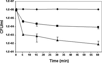
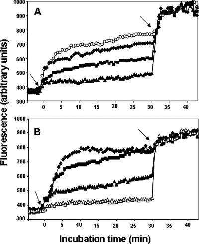
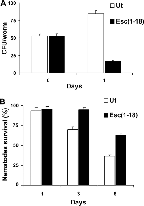
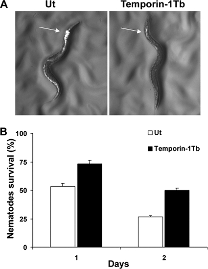
The emergence of multidrug-resistant (MDR) microorganisms makes it increasingly difficult to treat infections. These infections include those associated with Pseudomonas aeruginosa, which are hard to eradicate, especially in patients with a compromised immune system. Naturally occurring membrane-active cationic antimicrobial peptides (CAMPs) serve as attractive candidates for the development of new therapeutic agents. Amphibian skin is one of the richest sources for such peptides, but only a few studies on their in vivo activities and modes of action have been reported. We investigated (i) the activity and mechanism underlying the killing of short CAMPs from frog skin (e.g., temporins and esculentin fragments) on an MDR clinical isolate of P. aeruginosa and (ii) their in vivo antibacterial activities and modes of action, using the minihost model of Caenorhabditis elegans. Our data revealed that in vivo, both temporin-1Tb and esculentin(1-18) were highly active in promoting the survival of Pseudomonas-infected nematodes, although temporin-1Tb did not show significant activity in vitro under the experimental conditions used. Importantly, esculentin(1-18) permeated the membrane of Pseudomonas cells within the infected nematode. To the best of our knowledge, this is the first report showing the ability of a CAMP to permeate the microbial membrane within a living organism. Besides shedding light on a plausible mode of action of frog skin CAMPs in vivo, our data suggest that temporins and esculentins would be attractive molecules as templates for the development of new therapeutics against life-threatening infections.


Figures

FIG. 1.
FIG. 1.
Time-kill curves of P. aeruginosa by esculentin fragments. Bacteria (1 × 10⁶ CFU/ml) were grown in MHB at 37°C, diluted in PBS, and incubated with Esc(1-18) (▪) and Esc(1-21) (▴) at the MIC (8 and 4 μM, respectively). Controls (⧫) were bacteria incubated in the presence of peptide solvent (20% ethanol) at a final concentration of 0.6%. The data are the means ± standard deviations (SD) of three independent experiments.
FIG. 2.
FIG. 2.
Effect of esculentin fragments on the IM permeation of P. aeruginosa. Cells (1 × 10⁵) were incubated in 100 μl of PBS with 1 μM Sytox Green. Once basal fluorescence reached a constant value, the peptide was added (left arrows; t = 0), and changes in fluorescence were monitored (λexc = 485 nm; λems = 535 nm) and plotted as arbitrary units. Maximal membrane permeation (right arrows; t = 30 min) was obtained after the addition of 1 mM EDTA plus 0.5% Triton X-100. The data points represent the means of triplicate samples, with SD values not exceeding 2.5% from a single experiment, representative of three different experiments. Peptide concentrations used for Esc(1-18) (A) and Esc(1-21) (B) were as follows: 1 μM (▵), 2 μM (▴), 4 μM (▪), 8 μM (⧫), and 16 μM (○). The fluorescence values of controls (bacteria without peptide) were subtracted from each sample.
FIG. 3.
FIG. 3.
In vivo toxicity assay of frog skin CAMPs on C. elegans worms and their efficacies on nematodes infected with an MDR strain of P. aeruginosa. (A) Adult C. elegans worms were incubated with different concentrations of CAMPs, as described in Materials and Methods. Nematode survival was monitored 48 h after peptide addition and is expressed as a percentage with respect to non-peptide-treated worms. The results are the means of two independent experiments; the error bars indicate SD. (B) Nematode infection was carried out for 6 h as described in Materials and Methods. C. elegans survival was monitored after 40 h of exposure to peptides in a liquid medium assay. Peptides were used at the following concentrations: Esc(1-18), 0.3 nM; Esc(1-21), 0.3 nM; temporin-1Tb, 0.3 nM; temporin-1Tf, 0.01 nM; and temporin-1Tl, 0.01 nM. Infected animals not treated with the peptides (Ut) were included for comparison. Control animals not exposed to the pathogen and fed with E. coli OP50 exhibited 100% viability. The results represent the means of at least three independent experiments; the error bars indicate SD.
FIG. 4.
FIG. 4.
Effect of Esc(1-18) on the number of live Pseudomonas cells within the worm gut and the animal's survival. Adult worms were infected with the MDR strain of P. aeruginosa for 6 h and then transferred to NGM agar plates supplemented with E. coli OP50, to which a solution of 0.5 nM Esc(1-18) was directly added. (A) CFU were determined in the guts of 10 live worms removed from the plates before (0 days) and after 1 day of peptide treatment. Infected nematodes not treated with the peptide (Ut) were included for comparison. (B) Survival of infected C. elegans upon peptide treatment was analyzed at the indicated time points in comparison to untreated infected nematodes (Ut). Control worms not exposed to the pathogen and fed with E. coli OP50 showed 100% viability at each time point. The reported values represent the means of at least three independent experiments; the error bars indicate SD.
FIG. 5.
FIG. 5.
Effect of temporin-1Tb on the colonization of P. aeruginosa ATCC 15692 within the nematode gut and the animal's survival. Nematodes were infected with the GFP-expressing P. aeruginosa strain ATCC 15692 for 16 h to increase the number of intestinal bacteria, as described in Materials and Methods. (A) Fluorescence photomicrographs of a representative nematode after 90 min of treatment with 0.5 nM temporin-1Tb. A representative non-peptide-treated animal (Ut) was included for comparison. The arrows indicate the anterior intestine. (B) Survival of infected nematodes after 1 and 2 days of treatment with temporin-1Tb at 0.5 nM in an agar medium assay in comparison with untreated infected nematodes (Ut). The reported values represent the means of at least three independent experiments; the error bars indicate SD. Control animals not exposed to the pathogen and fed with E. coli OP50 exhibited 100% viability at each time point. Similar data were obtained when temporin-1Tb was replaced by Esc(1-18) and therefore are not shown.
FIG. 6.
FIG. 6.
Effect of Esc(1-18) on the membrane permeation of the MDR strain of P. aeruginosa within nematodes. Infected nematodes were transferred to a 24-well microtiter plate (200 worms per well), each well containing 300 μl of K medium, and incubated with 1 μM Sytox Green as described in Materials and Methods. Once basal fluorescence reached a constant value (time zero), 10 nM Esc(1-18) was added, and changes in fluorescence intensity were monitored (λexc = 485 nm; λems = 535 nm) until the fluorescent signal reached a constant value (approximately 40 min). The increase in fluorescence reflected damage at the IM level of Pseudomonas cells. Infected nematodes not treated with the peptide, as well as peptide-treated uninfected worms, were used for comparison. The reported values represent the means of two independent experiments; the error bars indicate SD.

References

    1. Ausubel, F. M. 2005. Are innate immune signaling pathways in plants and animals conserved? Nat. Immunol. 6:973-979. - PubMed
    1. Bhavsar, A. P., and E. D. Brown. 2006. The worm turns for antimicrobial discovery. Nat. Biotechnol. 24:1098-1100. - PubMed
    1. Carotenuto, A., S. Malfi, M. R. Saviello, P. Campiglia, I. Gomez-Monterrey, M. L. Mangoni, L. M. Gaddi, E. Novellino, and P. Grieco. 2008. A different molecular mechanism underlying antimicrobial and hemolytic actions of temporins A and L. J. Med. Chem. 51:2354-2362. - PubMed
    1. Chamilos, G., M. S. Lionakis, R. E. Lewis, and D. P. Kontoyiannis. 2007. Role of mini-host models in the study of medically important fungi. Lancet Infect. Dis. 7:42-55. - PubMed
    1. Cirioni, O., C. Silvestri, R. Ghiselli, F. Orlando, A. Riva, F. Mocchegiani, L. Chiodi, S. Castelletti, E. Gabrielli, V. Saba, G. Scalise, and A. Giacometti. 2008. Protective effects of the combination of alpha-helical antimicrobial peptides and rifampicin in three rat models of Pseudomonas aeruginosa infection. J. Antimicrob. Chemother. 62:1332-1338. - PubMed

Publication types

MeSH terms

LinkOut - more resources