. 2010 Oct 14;116(15):2826-35.
doi: 10.1182/blood-2010-03-275776. Epub 2010 Jul 6.

A transgenic mouse model demonstrates a dominant negative effect of a point mutation in the RPS19 gene associated with Diamond-Blackfan anemia

Affiliations

A transgenic mouse model demonstrates a dominant negative effect of a point mutation in the RPS19 gene associated with Diamond-Blackfan anemia

Emily E Devlin et al. Blood. .

Abstract

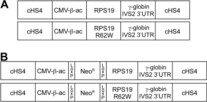
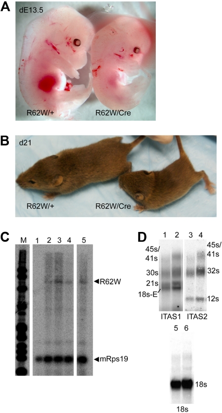
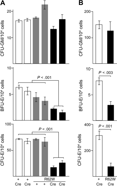
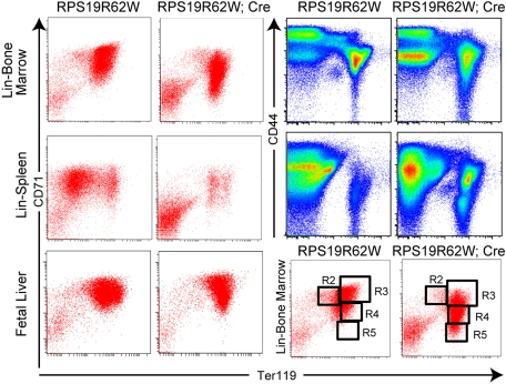
Diamond Blackfan anemia (DBA) is an inherited erythroblastopenia associated with mutations in at least 8 different ribosomal protein genes. Mutations in the gene encoding ribosomal protein S19 (RPS19) have been identified in approximately 25% of DBA families. Most of these mutations disrupt either the translation or stability of the RPS19 protein and are predicted to cause DBA by haploinsufficiency. However, approximately 30% of RPS19 mutations are missense mutations that do not alter the stability of the RPS19 protein and are hypothesized to act by a dominant negative mechanism. To formally test this hypothesis, we generated a transgenic mouse model expressing an RPS19 mutation in which an arginine residue is replaced with a tryptophan residue at codon 62 (RPS19R62W). Constitutive expression of RPS19R62W in developing mice was lethal. Conditional expression of RPS19R62W resulted in growth retardation, a mild anemia with reduced numbers of erythroid progenitors, and significant inhibition of terminal erythroid maturation, similar to DBA. RNA profiling demonstrated more than 700 dysregulated genes belonging to the same pathways that are disrupted in RNA profiles of DBA patient cells. We conclude that RPS19R62W is a dominant negative DBA mutation.


Figures

Figure 1
Figure 1
Diagrams of the constructs used to generate transgenic mice. All constructs are flanked by the chicken β-globin HS4 insulator (cHS4). The CMV enhancer/chicken β-actin promoter (CMV-β-ac) is used to express either the wild-type RPS19 cDNA (RPS19) or an RPS19 cDNA with the R62W point mutation (RPS19R62W). After the RPS19 stop codon, the 3′ end of the human γ-globin gene, including 52 bp of exon 2 (out of frame), IVS2, exon 3, and the 3′untranslated region (γ-globin IVS2 3′LTR) are attached to provide sequences for splicing, poly A addition, and mRNA stability. (A) Constructs that allow constitutive expression of the RPS19 transgenes. (B) Constructs that allow conditional expression of the RPS19 transgenes after Cre-mediated excision of the PGK-Neo sequences (NeoR) flanked by Lox P sites that were inserted between the CVM enhancer/chicken β-actin promoter (CMV-β-ac) and RPS19 cDNAs.
Figure 2
Figure 2
Analysis of transgenic mice carrying the conditional RPS19R62W construct with (R62W/Cre) and without (R62W/+) the Prion-Cre transgene. (A) Day 13.5 embryos. (B) Three-week-old pups. (C) RNase protection analysis of fetal liver RNA from day 13.5 embryos. Lane 1 indicates RNA from R62W/+ embryo; and lanes 2 to 5, RNA from R62W/Cre embryos. The transgenic RPS19 mRNA protects a 584-bp fragment of the probe. The mouse Rps19 mRNA protects 2 93-bp fragments of the probe, providing an internal control. (D) Northern blot analysis of rRNA processing. The individual rRNA products are indicated at the side of the figure. Lanes 1 and 2 indicate bone marrow RNA from R62W/+ (lane 1) R62W/Cre (lane 2) animals probed with an ITS1 probe; lanes 3 and 4, bone marrow RNA from R62W/+ (lane 3) R62W/Cre (lane 4) animals probed with an ITS2 probe; lanes 5 and 6, bone marrow RNA from R62W/+ (lane 5) R62W/Cre (lane 6) animals probed with an 18s rRNA probe. The apparent increases in the level of 21s (ITS1) and 12s (ITS2) are the result of the increased amount of R62W/Cre RNA in lanes 2, 4, and 6 as demonstrated by the level of 18s hybridization.
Figure 3
Figure 3
Analysis of hematopoietic colony formation in RPS19R62W transgenic mice. (A) Analysis of CFU-GM, BFU-E, and CFU-E from individual day 13.5 fetal livers. Cells were plated in triplicate. The genotypes of the embryos appear below the bars. The Prion-Cre transgene alone = + Cre (□); the R62W transgene alone = Tg + (□); the R62W transgene plus Prion-Cre = Tg Cre (■). (B) Analysis of CFU-GM, BFU-E, and CFU-E from 4- to 6-week-old mice. The data are compiled from 4 to 6 individual mice; cells from each mouse were plated in duplicate. The genotypes of the embryos appear below the bars. The Prion-Cre transgene alone = + Cre (□); the R62W transgene plus Prion-Cre = Tg Cre (■).
Figure 4
Figure 4
FACS analysis of erythropoiesis in R62W transgenic mice. (Left) Analysis of Lin bone marrow cells (top) Lin spleen cells (center) and fetal liver cells (bottom), stained with antibodies against CD71 and Ter119. (Right) Analysis Lin bone marrow (top) and Lin spleen cells (center) stained with antibiodies against CD44 and Ter119. (Bottom right) Gates used for sorting. Cells from mice carrying the RPS19R62W transgene alone = RPS19R62W; cells from mice carrying the RPS19R62W transgene and the Prion-Cre transgene = RPS19R62W; Cre.
Figure 5
Figure 5
Morphologic analysis of Wright-Giemsa-stained Lin bone marrow cells from R62W transgenic mice in the basophilic erythroblast (R3) and reticulocyte (R5) gates. The analysis was performed on a Nikon Eclipse TE300 microscope with a 40×/0.75 NA oil objective under Cargill Immersion oil (type 5). Images were taken with a Nikon N70 camera. No subsequent manipulations were performed. (Left) Analysis of cells from mice carrying the RPS19R62W transgene alone (RPS19R62W). (Right) Analysis of cells from mice carrying the RPS19R62W transgene and the Prion-Cre transgene (RPS19R62W; Cre). Note the binucleate cells among the RPS19R62W, Cre basophilic erythroblasts (R3), and the poorly hemoglobinized and nucleated cells among the RPS19R62W, Cre reticulocytes (R5).
Figure 6
Figure 6
Cell-cycle analysis of erythroid progenitors (R2) and basophilic erythroblasts (R3) in day 13.5 fetal liver cells from R62W transgenic mice. (A) Cell-cycle analysis of fetal liver R2 cells from embryos carrying the RPS19R62W transgene alone (□), and from embryos carrying the RPS19R62W transgene and the Prion-Cre transgene (■). (B) Cell-cycle analysis of fetal liver R3 cells from embryos carrying the RPS19R62W transgene alone (□), and from embryos carrying the RPS19R62W transgene and the Prion-Cre transgene (■). The percentage of cells in each phase of the cell cycle is indicated on the y-axis, and the cell-cycle phases (G1, S, G2/M) are on the x-axis. Bars represent data from 4 to 6 individual embryos. (C) Cell-cycle profile of fetal liver R3 cells from embryos carrying the RPS19R62W transgene alone. (D) Cell-cycle profile of fetal liver R3 cells from embryos carrying the RPS19R62W transgene and the Prion-Cre transgene.

Comment in

Similar articles

Cited by

References

    1. Josephs HW. Anemia of infancy and early childhood. Medicine. 1936;15:307–451.
    1. Diamond LK, Blackfan KD. Hypoplastic anemia. Am J Dis Child. 1938;56:464–467.
    1. Willig TN, Niemeyer CM, Leblanc T, et al. Identification of new prognosis factors from the clinical and epidemiologic analysis of a registry of 229 Diamond-Blackfan anemia patients: DBA group of Société d'Hématologie et d'Immunologie pédiatrique (SHIP), Gesellshaft für Pädiatrische Onkologie und Hämatologie (GPOH), and the European Society for Pediatric Hematology and Immunology (ESPHI). Pediatr Res. 1999;46(5):553–561. - PubMed
    1. Ramenghi U, Garelli E, Valtolina S, et al. Diamond-Blackfan anemia in the Italian population. Br J Haematol. 1999;104(4):841–848. - PubMed
    1. Ball SE, McGuckin CP, Jenkins G, Gordon-Smith EC. Diamond-Blackfan anemia in the UK: analysis of 80 cases from a 20-year birth cohort. Br J Haematol. 1996;94(4):645–653. - PubMed

Publication types