Crystallization and preliminary X-ray diffraction analysis of the N-terminal domain of human coronavirus OC43 nucleocapsid protein
- PMID: 20606281
- PMCID: PMC2898469
- DOI: 10.1107/S1744309110017616
Crystallization and preliminary X-ray diffraction analysis of the N-terminal domain of human coronavirus OC43 nucleocapsid protein
Abstract
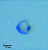
The N-terminal domain of nucleocapsid protein from human coronavirus OC43 (HCoV-OC43 N-NTD) mostly contains positively charged residues and has been identified as being responsible for RNA binding during ribonucleocapsid formation in the coronavirus. In this study, the crystallization and preliminary crystallographic analysis of HCoV-OC43 N-NTD (amino acids 58-195) with a molecular weight of 20 kDa are reported. HCoV-OC43 N-NTD was crystallized at 293 K using PEG 1500 as a precipitant and a 99.9% complete native data set was collected to 1.7 A resolution at 100 K with an overall R(merge) of 5.0%. The crystals belonged to the hexagonal space group P6(5), with unit-cell parameters a = 81.57, c = 42.87 A. Solvent-content calculations suggest that there is likely to be one subunit of N-NTD in the asymmetric unit.
Figures


Similar articles
-
Crystal structure-based exploration of the important role of Arg106 in the RNA-binding domain of human coronavirus OC43 nucleocapsid protein.Biochim Biophys Acta. 2013 Jun;1834(6):1054-62. doi: 10.1016/j.bbapap.2013.03.003. Epub 2013 Mar 15. Biochim Biophys Acta. 2013. PMID: 23501675 Free PMC article.
-
Immunoreactivity characterisation of the three structural regions of the human coronavirus OC43 nucleocapsid protein by Western blot: implications for the diagnosis of coronavirus infection.J Virol Methods. 2013 Feb;187(2):413-20. doi: 10.1016/j.jviromet.2012.11.009. Epub 2012 Nov 19. J Virol Methods. 2013. PMID: 23174159 Free PMC article.
-
Crystallographic analysis of the N-terminal domain of Middle East respiratory syndrome coronavirus nucleocapsid protein.Acta Crystallogr F Struct Biol Commun. 2015 Aug;71(Pt 8):977-80. doi: 10.1107/S2053230X15010146. Epub 2015 Jul 28. Acta Crystallogr F Struct Biol Commun. 2015. PMID: 26249685 Free PMC article.
-
Elucidation of the stability and functional regions of the human coronavirus OC43 nucleocapsid protein.Protein Sci. 2009 Nov;18(11):2209-18. doi: 10.1002/pro.225. Protein Sci. 2009. PMID: 19691129 Free PMC article.
-
[Molecular epidemiological study of human coronavirus OC43 in Shanghai from 2009-2016].Zhonghua Yu Fang Yi Xue Za Zhi. 2018 Jan 6;52(1):55-61. doi: 10.3760/cma.j.issn.0253-9624.2018.01.011. Zhonghua Yu Fang Yi Xue Za Zhi. 2018. PMID: 29334709 Chinese.
Cited by
-
Crystal structure-based exploration of the important role of Arg106 in the RNA-binding domain of human coronavirus OC43 nucleocapsid protein.Biochim Biophys Acta. 2013 Jun;1834(6):1054-62. doi: 10.1016/j.bbapap.2013.03.003. Epub 2013 Mar 15. Biochim Biophys Acta. 2013. PMID: 23501675 Free PMC article.
-
Structural basis for the identification of the N-terminal domain of coronavirus nucleocapsid protein as an antiviral target.J Med Chem. 2014 Mar 27;57(6):2247-57. doi: 10.1021/jm500089r. Epub 2014 Mar 12. J Med Chem. 2014. PMID: 24564608 Free PMC article.
-
Human Coronavirus OC43 as a Low-Risk Model to Study COVID-19.Viruses. 2023 Feb 20;15(2):578. doi: 10.3390/v15020578. Viruses. 2023. PMID: 36851792 Free PMC article. Review.
-
Exploring beyond clinical routine SARS-CoV-2 serology using MultiCoV-Ab to evaluate endemic coronavirus cross-reactivity.Nat Commun. 2021 Feb 19;12(1):1152. doi: 10.1038/s41467-021-20973-3. Nat Commun. 2021. PMID: 33608538 Free PMC article.
-
Immunoreactivity characterisation of the three structural regions of the human coronavirus OC43 nucleocapsid protein by Western blot: implications for the diagnosis of coronavirus infection.J Virol Methods. 2013 Feb;187(2):413-20. doi: 10.1016/j.jviromet.2012.11.009. Epub 2012 Nov 19. J Virol Methods. 2013. PMID: 23174159 Free PMC article.
References
-
- Collaborative Computational Project, Number 4 (1994). Acta Cryst. D50, 760–763. - PubMed
Publication types
MeSH terms
Substances
LinkOut - more resources
Full Text Sources
Research Materials

