Review
. 2010;92(2):81-99.
doi: 10.1159/000314193. Epub 2010 Jul 7.

Human GnRH deficiency: a unique disease model to unravel the ontogeny of GnRH neurons

Affiliations
Review

Human GnRH deficiency: a unique disease model to unravel the ontogeny of GnRH neurons

Ravikumar Balasubramanian et al. Neuroendocrinology. 2010.

Abstract

Evolutionary survival of a species is largely a function of its reproductive fitness. In mammals, a sparsely populated and widely dispersed network of hypothalamic neurons, the gonadotropin-releasing hormone (GnRH) neurons, serve as the pilot light of reproduction via coordinated secretion of GnRH. Since it first description, human GnRH deficiency has been recognized both clinically and genetically as a heterogeneous disease. A spectrum of different reproductive phenotypes comprised of congenital GnRH deficiency with anosmia (Kallmann syndrome), congenital GnRH deficiency with normal olfaction (normosmic idiopathic hypogonadotropic hypogonadism), and adult-onset hypogonadotropic hypogonadism has been described. In the last two decades, several genes and pathways which govern GnRH ontogeny have been discovered by studying humans with GnRH deficiency. More importantly, detailed study of these patients has highlighted the emerging theme of oligogenicity and genotypic synergism, and also expanded the phenotypic diversity with the documentation of reversal of GnRH deficiency later in adulthood in some patients. The underlying genetic defect has also helped understand the associated nonreproductive phenotypes seen in some of these patients. These insights now provide practicing clinicians with targeted genetic diagnostic strategies and also impact on clinical management.


Figures

Fig. 1
Fig. 1
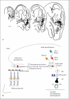
GnRH migration. a Migratory route of GnRH neurons in the embryonic mouse brain. GnRH neurons (black dots) originate in the medial wall of the olfactory placode, migrate along the olfactory axons and enter the brain perforating the cribriform plate. They then migrate to reach their final destination in the preoptic area (poa) of the hypothalamus. GnRH neurons on embryonic day 11 are seen in the vomeronasal organ (vno) and medial wall of the olfactory placode. In the 16-day-old fetal mouse brain, most of the GnRH neurons have reached the hypothalamus. gt = Ganglion terminale; ob = olfactory bulb. Reproduced with permission from Schwanzel-Fukuda and Pfaff [8]. b Cartoon of GnRH migration in humans. GnRH neurons originate in the embryonic olfactory placode and migrate along olfactory axons, penetrate the cribriform plate and migrate across the olfactory bulb, aided by various chemoattractive and repulsion factors and eventually reach the preoptic area of the hypothalamus, where they synchronize pulsatile GnRH secretion. Kisspeptin (KISS1) and the tachykinin (TAC) neurons serve as upstream modulators of GnRH secretion. Upon GnRH stimulation, the pituitary (PIT) secretes LH and follicle-stimulating hormone (FSH), which in turn regulate gonadal steroidogenesis and gametogenesis.
Fig. 2
Fig. 2
GnRH pulsatile secretion in normal puberty and disorders of pubertal development. a Pulsatile secretion of GnRH is fully active during the early neonatal period (‘mini-puberty of infancy’), followed by quiescence during childhood, and reactivates in adolescence signalling normal puberty. b Delayed activation of the GnRH pulse generator results in delayed puberty. c Premature activation of the GnRH pulse generator in early childhood results in precocious puberty.
Fig. 3
Fig. 3
KS and nIHH. a KS results from a developmental defect in olfactory bulb (OB) morphogenesis and GnRH neuronal migration resulting in failure of GnRH neurons to arrive at the preoptic area (POA) in the hypothalamus and failure of GnRH secretion. The GnRH neurons are arrested in their extracerebral route. Mutations in KAL1, PROK2, PROKR2, FGFR1, FGF8, NELF and CHD7 have been associated with KS. b nIHH results from either defective pulsatile secretion of GnRH or defective GnRH action at the pituitary. Mutations in GNRH1, KISS1R, GNRHR, PROK2, PROKR2, FGFR1, TAC3, TACR3, CHD7 and NROB1 genes have been associated with nIHH.
Fig. 4
Fig. 4
Phenotypic heterogeneity in human GnRH deficiency. GnRH deficiency can either be complete (a) or partial (b). c Some patients develop GnRH deficiency in adulthood following normal GnRH activation in the neonatal period (AHH). d Occasionally, following complete or partial GnRH deficiency, GnRH pulse generator activates in adulthood (reversal of GnRH deficiency).
Fig. 5
Fig. 5
Genetics of puberty. The precise genetic basis of the extreme tails of normal puberty is unclear. The genetic variants causing GnRH deficiency and precocious puberty may possibly explain the heritability of early and delayed puberty in the normal population.
Fig. 6
Fig. 6
KAL1 mutations in human GnRH deficiency. a Gene deletions (black lines) in KAL1 gene causing KS. b Schematic of exons of KAL1 gene. c Schematic of the anosmin-1 protein displaying known point mutations (in hexagons) causing KS. Anosmin-1, a multidomain protein, consists of a N-terminal signal peptide followed by a cysteine-rich region (CYS), a WAP-like four-disulfide core motif, four tandem FnIII repeats and a C-terminal histidine-rich region (H) (insertions, deletions, duplications and intronic changes not shown).
Fig. 7
Fig. 7
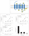
KISS1R and human GnRH deficiency. a Schematic of the predicted KISS1R protein displaying known mutations causing nIHH in humans. b Dose-response curves for the ligand-stimulated production of inositol phosphate in KISS1R mutant constructs, corrected for protein content. i Curve for the L148S mutation (three independent experiments, each performed in triplicate). ii Curve for the R331X mutation (two independent experiments, each performed in triplicate). iii Curve for the X399R polyA stop mutation (two independent experiments, each performed in triplicate). The percentages on the y-axis represent the percentages of the maximal stimulation for each GPR54 construct. iv The relative quantification of the wild-type and mutant GPR54 allele expression in lymphoblastoid cell lines as measured by quantitative RT-PCR. Reproduced with permission from Seminara et al. [50].
Fig. 8
Fig. 8
FGF pathway in human GnRH deficiency. a Schematic of FGFR1 protein displaying mutations causing KS (blue) and nIHH (black). The extracellular domain of FGFR1 contains a signal peptide (SP), three extracellular immunoglobulin-like (D1, D2, and D3) domains, followed by the TM, and an intracellular domain comprising two tyrosine kinase subdomains (PTK). The acidic box (AB) and the docking protein FRS2 domain in the extracellular domain and intracellular domain, respectively, are specific features of FGF receptors. b Genomic structure and differential splicing of the human FGF8 gene. i Structure of the FGF8 gene. Boxes denote exons; lines denote introns. ii Schematic of the four FGF8 isoforms identified in humans with mutations identified to date are indicated by arrows and numbered according to the FGF8f and FGF8b protein isoforms (∗ homozygous mutations). Reproduced from Falardeau et al. [71].
Fig. 9
Fig. 9
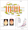
Prokineticin 2 pathway and human GnRH deficiency. a Schematic of the predicted PROKR2 protein displaying mutations causing KS (yellow boxes) and nIHH (white boxes). b Morphological examination of wild-type PROK2+/+ and PROK2–/– mice showing hypoplasia of olfactory bulbs (red circle). Reproduced with permission from Ng et al. [79]. c In vitro activity of a selection of PROKR2 mutants in an intracellular Ca2+ assay. Reproduced with permission from Cole et al. [90].

References

    1. Wray S, Hoffman G. A developmental study of the quantitative distribution of LHRH neurons within the central nervous system of postnatal male and female rats. J Comp Neurol. 1986;252:522–531. - PubMed
    1. Merchenthaler I, et al. Gonadotropin-releasing hormone (GnRH) neurons and pathways in the rat brain. Cell Tissue Res. 1984;237:15–29. - PubMed
    1. Grumbach MM. A window of opportunity: the diagnosis of gonadotropin deficiency in the male infant. J Clin Endocrinol Metab. 2005;90:3122–3127. - PubMed
    1. Conn PM, Crowley WF., Jr Gonadotropin-releasing hormone and its analogs. Annu Rev Med. 1994;45:391–405. - PubMed
    1. Antunes JL, et al. Luteinizing hormone-releasing hormone in human pituitary blood. J Neurosurg. 1978;49:382–386. - PubMed

MeSH terms