. 2010 Aug 18;29(16):2841-57.
doi: 10.1038/emboj.2010.143. Epub 2010 Jul 6.

ALS-associated fused in sarcoma (FUS) mutations disrupt Transportin-mediated nuclear import

Affiliations

ALS-associated fused in sarcoma (FUS) mutations disrupt Transportin-mediated nuclear import

Dorothee Dormann et al. EMBO J. .

Abstract

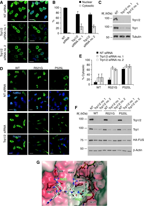
Mutations in fused in sarcoma (FUS) are a cause of familial amyotrophic lateral sclerosis (fALS). Patients carrying point mutations in the C-terminus of FUS show neuronal cytoplasmic FUS-positive inclusions, whereas in healthy controls, FUS is predominantly nuclear. Cytoplasmic FUS inclusions have also been identified in a subset of frontotemporal lobar degeneration (FTLD-FUS). We show that a non-classical PY nuclear localization signal (NLS) in the C-terminus of FUS is necessary for nuclear import. The majority of fALS-associated mutations occur within the NLS and impair nuclear import to a degree that correlates with the age of disease onset. This presents the first case of disease-causing mutations within a PY-NLS. Nuclear import of FUS is dependent on Transportin, and interference with this transport pathway leads to cytoplasmic redistribution and recruitment of FUS into stress granules. Moreover, proteins known to be stress granule markers co-deposit with inclusions in fALS and FTLD-FUS patients, implicating stress granule formation in the pathogenesis of these diseases. We propose that two pathological hits, namely nuclear import defects and cellular stress, are involved in the pathogenesis of FUS-opathies.


Conflict of interest statement

The authors declare that they have no conflict of interest.

Figures

Figure 1
Figure 1
The C-terminal tail of FUS (FUS514−526) is necessary and sufficient for nuclear import. (A) Schematic diagram of the domain structure of FUS. Mutations identified in fALS patients are shown below; 12 out of the 22 known mutations are clustered in the C-terminal tail (residues 514–526). (B) Alignment of the FUS C-termini of different species shows that the sequence of this domain is well conserved during evolution (identical residues are highlighted in yellow, homologous residues in light grey). (C) N-terminally HA-tagged wild-type (WT) FUS and the indicated point mutants were transiently expressed in HeLa cells; 24 h post-transfection cells were stained with an HA-specific antibody (green), a nuclear counter-stain (blue) and analysed by confocal microscopy. Although the WT protein is located almost exclusively in the nucleus, deletion of the C-terminal 13 amino acids (Δ514–526) or replacement of arginine residues by alanine (R521A/R522A/R524A or R514A/R518A) leads to a predominantly cytosolic localization. Scale bar, 20 μm. (D) Quantification of nuclear and cytosolic fluorescence intensities. Error bars indicate s.d. (E) The indicated sequences were fused to the C-terminus of the cytosolic reporter GST-GFP and reporter constructs were transiently expressed in HeLa cells; 24 h post-transfection cells were stained with a GFP-specific antibody (green) and a nuclear counter-stain (blue) and localization of the reporter proteins was analysed by confocal microscopy. Without active nuclear import, GST-GFP is localized predominantly in the cytosol (first panel), whereas attachment of the well-characterized NLS of the SV40 large T antigen (SV40-NLS) or the last 13 amino acids of FUS (FUS514−526) efficiently mediate nuclear import (second and third panel). The same amino acids arranged in random order (FUS514−526 scrambled) are not sufficient for mediating nuclear import (last panel). Scale bar, 20 μm. (F) Quantification of nuclear and cytosolic fluorescence intensities. Error bars indicate s.d.
Figure 2
Figure 2
fALS-associated mutations in important residues of the FUS NLS disrupt nuclear import. (A) HA-tagged wild-type (WT) FUS or FUS carrying the indicated C-terminal point mutations was transiently expressed in HeLa cells. Cells were stained with an HA-specific antibody (green) and a nuclear counter-stain (blue) and were analysed by confocal microscopy. R521G and R524S show a mild, R522G and P525L a strong cytosolic mislocalization, suggesting that these mutations disrupt important residues of the FUS NLS. Scale bar, 20 μm. (B) Quantification of nuclear and cytosolic fluorescence intensities. Error bars indicate s.d. The degree of cytoplasmic mislocalization inversely correlates with the age of onset of the individual point mutations. (C) HA-FUS protein levels in HeLa cells transiently transfected with the indicated HA-FUS constructs were analysed by immunoblotting with an HA-specific antibody (upper panel). LDH served as a loading control (lower panel). (D) HA-tagged wild-type (WT) FUS or FUS carrying the indicated N-terminal point mutations was transiently expressed in HeLa cells. Cells were stained with an HA-specific antibody (green) and a nuclear counter-stain (blue) and were analysed by confocal microscopy. The nuclear/cytosolic distribution of the N-terminal point mutants is indistinguishable from the WT protein. Scale bar, 20 μm. (E) Quantification of nuclear and cytosolic fluorescence intensities. Error bars indicate s.d.
Figure 3
Figure 3
The FUS-P525L mutation disrupts nuclear import in primary neurons and prevents import of a cytosolic reporter in vitro and in vivo. (A) Cultured neurons from E19 rat hippocampus or frontal cortex were transfected with HA-tagged FUS-WT or P525L, mCherry (red) was co-expressed to visualize neuron morphology. Two days post-transfection, cells were stained with an HA-specific antibody (green) and a nuclear counter-stain (blue) and were analysed by confocal microscopy. FUS-WT is mostly confined to the nucleus, whereas the P525L mutant shows abundant staining in the whole cell body and in neurites. Scale bar, 20 μm. (B) Quantification of nuclear and cytosolic fluorescence intensities. Z-stacks were taken and all planes were projected into a single image along the z axis (maximal projection). Ten fields were analysed for each sample and mean values were calculated. Error bars indicate s.d. (C) To confirm that fALS-associated point mutations disrupt nuclear import, the P525L mutation was introduced into the GST-GFP-FUS514−526 reporter and was analysed for its effect on nuclear import activity. Localization of the P525L-containing reporter is identical to that of the FUS514−526 scrambled reporter, showing that this point mutation completely disrupts activity of the C-terminal NLS. Scale bar, 20 μm. (D) Quantification of nuclear and cytosolic fluorescence intensities. Error bars indicate s.d. (E) To confirm functionality of the FUS-NLS in vivo, the indicated GST-GFP reporter constructs were injected into fertilized zebrafish eggs. On day 2 post-fertilization, embryos were stained with a GFP-specific antibody (green) and a nuclear counter-stain (blue), and subcellular localization of the reporter constructs was analysed in muscle cells and spinal cord neurons by confocal microscopy. In both cell types, the FUS514−526 WT sequence mediates efficient nuclear import (left panels), whereas reporter proteins carrying the P525L mutation or scrambled NLS remain cytosolic (middle and right panels). Arrowheads indicate axonal localization of reporter proteins in spinal cord neurons. Scale bar, 10 μm.
Figure 4
Figure 4
Transportin is required for nuclear import of FUS. (A) The two Trp homologues, Trp1 and Trp2, were silenced by siRNA-mediated knockdown, using two different siRNA pools (no. 1 and no. 2). A non-targeting (NT) siRNA was used as a negative control; 72 h post-transfection, cells were stained with an FUS-specific antibody (green) and a nuclear counter-stain (blue) and were analysed by confocal microscopy. Trp1/2 double knockdown leads to a partial cytoplasmic redistribution, showing that Trp is involved in nuclear import of FUS. Scale bar, 20 μm. (B) Quantification of nuclear and cytosolic fluorescence intensities. Error bars indicate s.d. (C) Verification of knockdown efficiency by immunoblot. Total cell lysates were examined with a pan-Trp (Trp1/2)- and a Trp1-specific antibody (upper two panels). α-Tubulin served as a loading control (lower panel). Note that the Trp1-specific antibody is more sensitive than the pan-Trp antiserum and detects residual levels of Trp1 (middle panel). (D) HeLa cells were transfected with NT siRNA or Trp1/2-specific siRNA pool no. 1 or no. 2 and 24 h later with the indicated HA-tagged FUS constructs. Another 24 h later, cells were stained with an HA-specific antibody (green) and a nuclear counter-stain (blue) and were analysed by confocal microscopy. Trp silencing leads to a dramatic cytosolic mislocalization of the otherwise weakly mislocalized R521G mutant, but has almost no further effect on the already strongly mislocalized P525L mutant. Scale bar, 20 μm. (E) Quantification of nuclear and cytosolic fluorescence intensities. Error bars indicate s.d. (F) Verification of knockdown efficiency and expression of HA constructs by immunoblot. Total cell lysates were examined with antibodies specific for Trp1/2, Trp1 and HA (upper three panels). β-actin served as a loading control (lowest panel). (G) Model of the FUS PY-NLS (stick model with grey carbons, red oxygens, blue nitrogens, important amino-acid residues labelled in green) bound to the semitransparent electrostatic surface of Trp coloured according to its calculated negative (−25 e/kT, red) and positive (+25 e/kT, blue) electrostatic surface potential. Underlying amino-acid residues of special importance for binding of the FUS-NLS are depicted as stick model and labelled in black. Residues responsible for the charged H-bond/salt-bridge contact to FUS-R522 and residues forming the hydrophobic pocked for FUS-PY526 and the H-bond network connecting to FUS-Y526 OH are shown with blue, orange and green carbons, respectively. H-bonds are indicated as broken black lines. This figure was made with pymol (DeLano Scientific LLC, USA, http://www.pymol.org).
Figure 5
Figure 5
Expression of a Trp-specific peptide inhibitor leads to the cytosolic redistribution of FUS, but not TDP-43. (A, B) A peptide competitor (M9M) designed to bind to the PY-NLS-binding site in Trp with very high affinity was expressed in primary rat cortical neurons (A) or HeLa cells (B) as a GFP-fusion protein (green). After staining with an FUS-specific antibody (red), cells were analysed by confocal microscopy. Expression of the Trp-specific inhibitor construct causes a marked cytoplasmic redistribution and localization of endogenous FUS in cytoplasmic punctate structures. Scale bar, 20 μm. Insert in (A) and panels on the right of (B) show magnifications of the boxed regions. (C) Quantification of the percentage of HeLa cells with exclusively nuclear, diffuse cytosolic and punctuate cytosolic FUS staining. Error bars indicate s.d. (D) To show selectivity of the M9M peptide inhibitor, GFP or GFP-M9M (green)-transfected HeLa cells were co-stained for endogenous FUS (red) and TDP-43 (white) and were analysed by confocal microscopy. In contrast to FUS, nuclear localization of TDP-43 is not affected by expression of the M9M construct. Scale bar, 20 μm.
Figure 6
Figure 6
Redistribution of FUS into cytoplasmic stress granules. (A) GFP-M9M (green)-transfected HeLa cells were co-stained for endogenous FUS (red) and the stress granule marker proteins TIAR, PABP-1, TIA-1, G3BP1 or the P body marker Dcp1 (white). Co-staining of FUS with TIAR, PABP-1, TIA-1 and G3BP1 shows that the punctate FUS-positive structures are stress granules. Note that there is no co-localization with the P body marker Dcp1. Scale bar, 20 μm. (B) GFP-M9M (green)-transfected HeLa cells were stained for endogenous FUS (red) and the stress granule marker G3BP1 (white). Where indicated, the polysome-stabilizing drug cycloheximide (CHX) was added for 1 h before fixation to prevent stress granule formation. Cycloheximide prevents formation of G3BP1- and FUS-positive cytosolic structures, confirming their stress granule identity. Scale bar, 20 μm.
Figure 7
Figure 7
Neuronal cytoplasmic inclusions (NCIs) in patients with FUS pathology contain the stress granule marker proteins PABP-1 and eIF4G. (A) Upper panels: PABP-1 immunohistochemistry performed on sections of post-mortem tissue reveals strongly immunoreactive NCIs in motor neurons in the spinal cord in fALS-R521C, in dentate granule cells of the hippocampus in aFTLD-U and NIFID as well as in motor neurons in the spinal cord in BIBD. In contrast, no PABP-1-labeled inclusions were detectable in dentate granule cells of the hippocampus in FTLD-TDP. Scale bar, 25 μm. Lower panels: double-label immunofluorescence stainings of the same cases and brain regions show co-localization of PABP-1 (red) with p62-positive inclusions (green) in fALS-R521C, aFTLD-U, NIFID and BIBD, but no PABP-1 staining in FTLD-TDP inclusions. Note that p62 is a robust marker of FUS and TDP-43 NCIs and was used because double labelling for FUS and PABP-1 was technically not possible, as available antibodies working on paraffin-embedded tissue were both rabbit polyclonal antisera. Scale bar, 12.5 μm. (B) eIF4G immunohistochemistry reveals labelling of NCIs in motor neurons in the spinal cord in fALS-R521C, in dentate granule cells of the hippocampus in aFTLD-U and NIFID and neurons in frontal cortex in BIBD. No NCIs were detectable in dentate granule cells in FTLD-TDP. Scale bar, 25 μm.
Figure 8
Figure 8
C-terminal fALS-associated FUS mutations favour recruitment of FUS into stress granules. (A) HeLa cells were transiently transfected with the indicated HA-tagged FUS constructs; 24 h post-transfection, cells were subjected to heat shock (44°C for 1 h, right panels) or were kept at control temperature (37°C, left panels). Cells were fixed, stained with an HA-specific antibody (green), a PABP-1-specific antibody (red) and a nuclear counter-stain (blue) and analysed by confocal microscopy. In contrast to WT-FUS, which remains almost exclusively nuclear on heat shock, all FUS mutants are recruited into PABP-1-positive stress granules. The amount of FUS in stress granules correlates with the cytoplasmic mislocalization and average age of disease onset of the individual point mutations, suggesting that cytoplasmic mislocalization favours recruitment of FUS to stress granules. Scale bar, 20 μm. (B) Primary rat hippocampal neurons were transiently transfected with HA-tagged FUS-WT or the P525L mutant, mCherry (red) was co-transfected to visualized neuron morphology. Two days post-transfection, cells were subjected to heat shock (44°C for 1 h) or were kept at control temperature (37°C) and were stained with an HA-specific antibody (green) and a TIAR-specific antibody (white). WT-FUS remains almost exclusively nuclear on heat shock, whereas the P525L mutant shows a mostly granular localization and co-localizes with TIAR-positive stress granules. Scale bar, 10 μm. (C) HeLa cells transiently transfected with the HA-tagged FUS-P525L mutant were subjected to heat shock (44°C for 1 h) or were kept at control temperature (37°C). Cells were stained with an HA-specific antibody (green), a TDP-43-specific antibody (red) and a nuclear counter-stain (blue) and analysed by confocal microscopy. TDP-43 is not recruited into FUS-P525L-containing stress granules. Scale bar, 20 μm.
Figure 9
Figure 9
A two hit model of FUS pathology. Green colour represents FUS distribution. For details see Discussion.

Comment in

References

    1. Anderson P, Kedersha N (2006) RNA granules. J Cell Biol 172: 803–808 - PMC - PubMed
    1. Anderson P, Kedersha N (2008) Stress granules: the Tao of RNA triage. Trends Biochem Sci 33: 141–150 - PubMed
    1. Andersson MK, Stahlberg A, Arvidsson Y, Olofsson A, Semb H, Stenman G, Nilsson O, Aman P (2008) The multifunctional FUS, EWS and TAF15 proto-oncoproteins show cell type-specific expression patterns and involvement in cell spreading and stress response. BMC Cell Biol 9: 37–53 - PMC - PubMed
    1. Arai T, Hasegawa M, Akiyama H, Ikeda K, Nonaka T, Mori H, Mann D, Tsuchiya K, Yoshida M, Hashizume Y, Oda T (2006) TDP-43 is a component of ubiquitin-positive tau-negative inclusions in frontotemporal lobar degeneration and amyotrophic lateral sclerosis. Biochem Biophys Res Commun 351: 602–611 - PubMed
    1. Ayala YM, Zago P, D'Ambrogio A, Xu YF, Petrucelli L, Buratti E, Baralle FE (2008) Structural determinants of the cellular localization and shuttling of TDP-43. J Cell Sci 121: 3778–3785 - PubMed

Publication types

MeSH terms