

Islet architecture: A comparative study
- PMID: 20606719
- PMCID: PMC2894473
- DOI: 10.4161/isl.1.2.9480
Islet architecture: A comparative study
Abstract
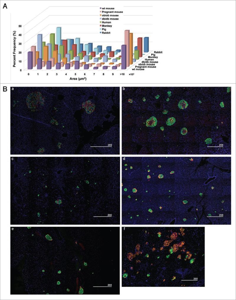
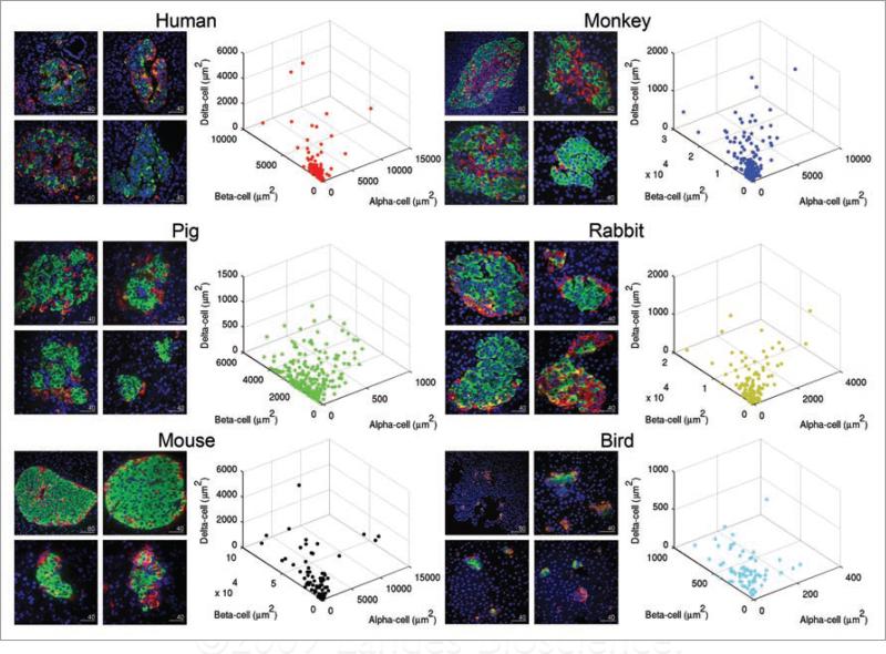
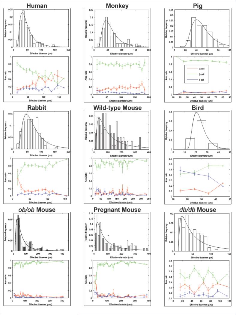
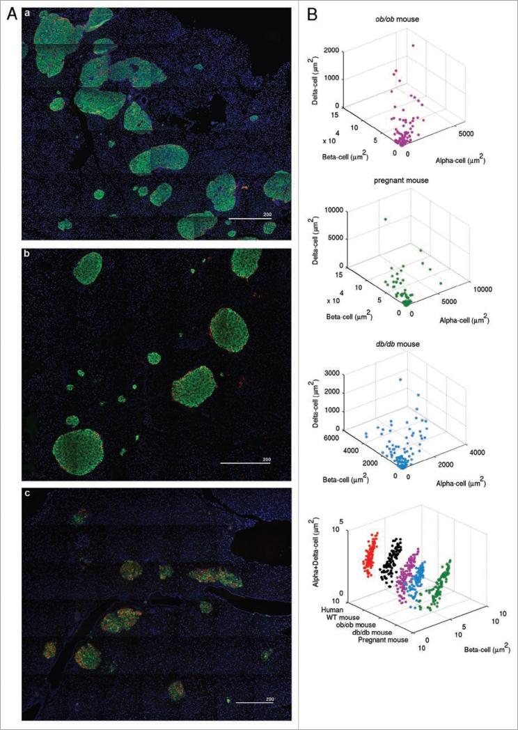
Emerging reports on the organization of the different hormone-secreting cell types (alpha, glucagon; beta, insulin; and delta, somatostatin) in human islets have emphasized the distinct differences between human and mouse islets, raising questions about the relevance of studies of mouse islets to human islet physiology. Here, we examine the differences and similarities between the architecture of human and mouse islets. We studied islets from various mouse models including ob/ob and db/db and pregnant mice. We also examined the islets of monkeys, pigs, rabbits and birds for further comparisons. Despite differences in overall body and pancreas size as well as total beta-cell mass among these species, the distribution of their islet sizes closely overlaps, except in the bird pancreas in which the delta-cell population predominates (both in singlets and clusters) along with a small number of islets. Markedly large islets (>10,000 mum(2)) were observed in human and monkey islets as well as in islets from ob/ob and pregnant mice. The fraction of alpha-, beta- and delta-cells within an islet varied between islets in all the species examined. Furthermore, there was variability in the distribution of alpha- and delta-cells within the same species. In summary, human and mouse islets share common architectural features that may reflect demand for insulin. Comparative studies of islet architecture may lead to a better understanding of islet development and function.
Keywords: diabetes; insulin resistance; pancreatic islets; pregnancy; α-cells; β-cells; δ-cells.
Figures
References
-
- Brissova M, Fowler MJ, Nicholson WE, Chu A, Hirshberg B, Harlan DM, et al. Assessment of human pancreatic islet architecture and composition by laser scanning confocal microscopy. J Histochem Cytochem. 2005;53:1087–97. - PubMed
-
- Orci L, Unger RH. Functional subdivision of islets of Langerhands and possible role of D Cells. Lancet. 1975;2:1243–4. - PubMed
-
- Samols E, Bonner-Weir S, Weir GC. Intra-islet insulin-glucagon-somatostatin relationships. Clin Endocrinol Metab. 1986;15:33–58. - PubMed
-
- Yukawa M, Takeuchi T, Watanabe T, Kitamura S. Proportions of Various Endocrine Cells in the Pancreatic Islets of Wood Mice. Anat Histol Embryol. 1999;28:13–6. - PubMed
Publication types
MeSH terms
Substances
Grants and funding
LinkOut - more resources
Full Text Sources
Other Literature Sources
Molecular Biology Databases
Miscellaneous