. 2009 Dec 1:1:15-32.
doi: 10.2147/ijicmr.s6776.

Cytokine-modified VSV is attenuated for neural pathology, but is both highly immunogenic and oncolytic

Affiliations

Cytokine-modified VSV is attenuated for neural pathology, but is both highly immunogenic and oncolytic

James Miller et al. Int J Interferon Cytokine Mediat Res. .

Abstract

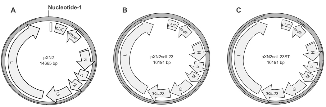
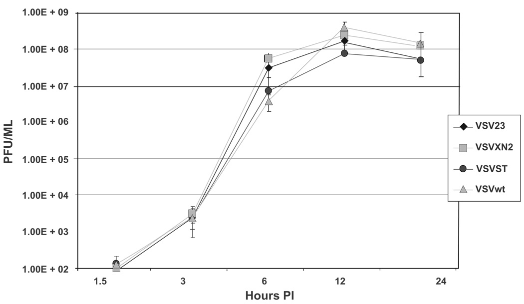
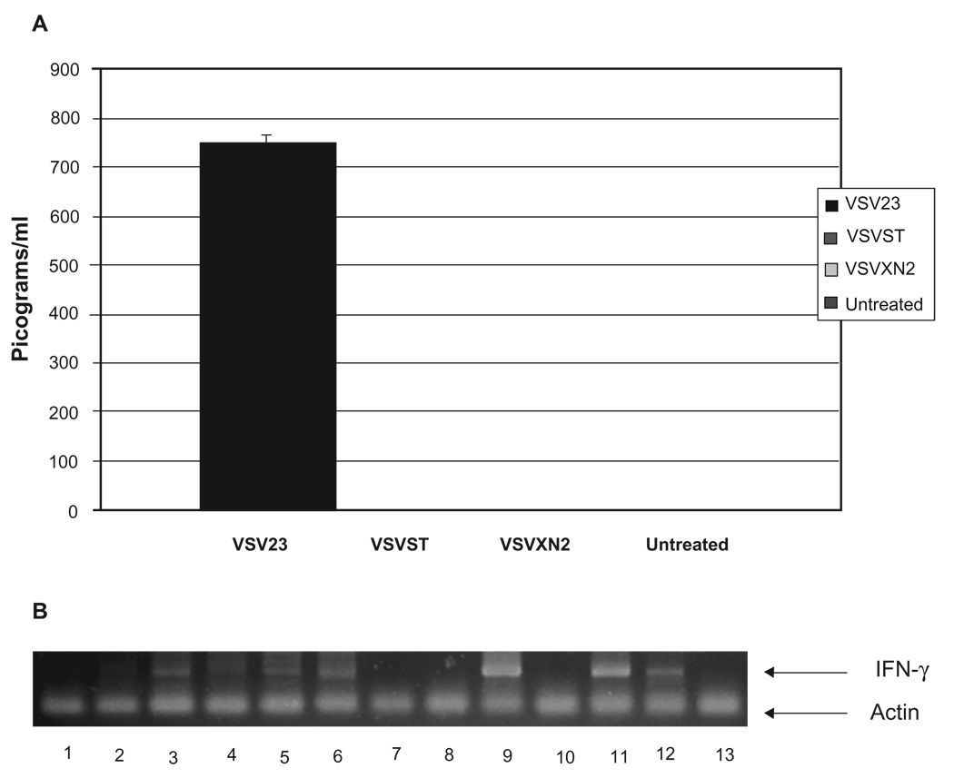
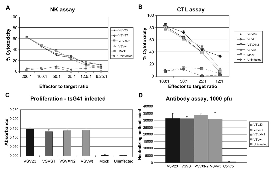
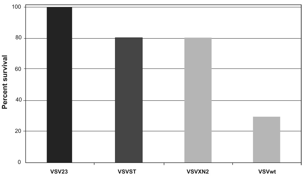
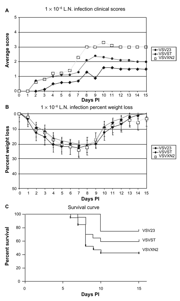
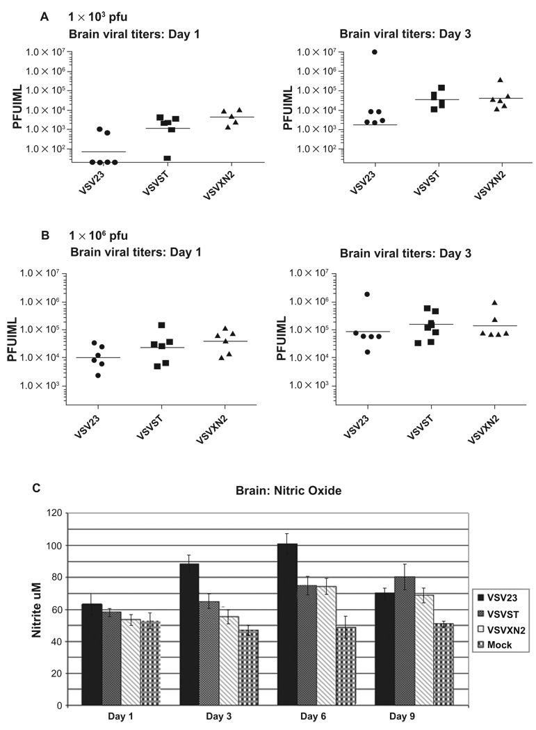
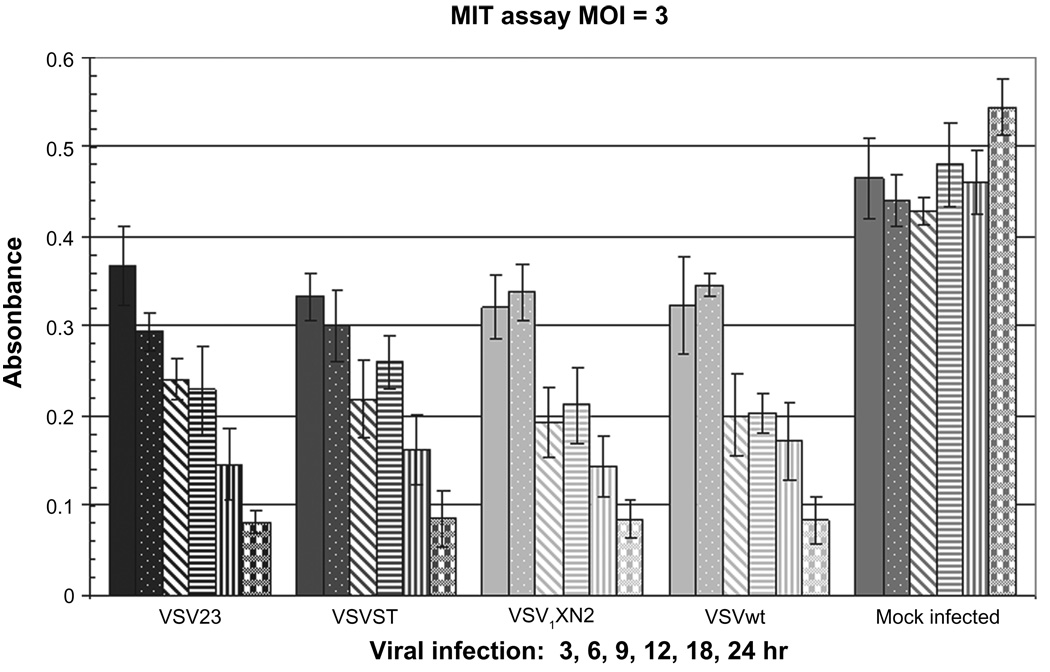
Vesicular stomatitis virus (VSV), an enveloped, nonsegmented, negative-stranded RNA virus, is being tested by several laboratories as an antitumor agent. Unfortunately, viral infection of the central nervous system (CNS) has been observed by many groups following administration to tumor-bearing animals. In rodents, VSV encephalitis is characterized by weight-loss, paralysis, and high mortality. In order to provide protection from VSV infection of the CNS after therapeutic administration, we have attenuated VSV by the introduction of the gene encoding the proinflammatory cytokine interleukin (IL)-23, and designated the new virus VSV23. We hypothesize that while VSV23 is replicating within tumors, resulting in tumor destruction, the expression of IL-23 will enhance host antitumor and antiviral immune responses. In the event that the virus escapes from the tumor, the host's immune system will be activated and the virus will be rapidly cleared from healthy tissue. Experimental VSV23 infection of the CNS is characterized by decreased viral replication, morbidity, and mortality. VSV23 is capable of stimulating the enhanced production of nitric oxide in the CNS, which is critical for elimination of VSV from infected neurons. Intraperitoneal administration of VSV23 stimulates both nonspecific natural killer cell, virus-specific cytolytic T lymphocyte and memory virus-specific proliferative T cell responses against wild-type VSV in splenocytes. Furthermore, VSV23 is able to replicate in, and induce apoptosis of tumor cells in vitro. These data indicate that VSV23 is immunogenic, attenuated and suitable for testing as an efficacious and safe oncolytic agent.


Figures

Figure 1
Figure 1
Virus production plasmids. A) VSVXN2. is a plasmid map of the 14.6 kb pXN2 plasmid that serves as the cloning vector in the recombinant VSV system and used for the rescue of VSVXN2. B) VSV23. A single chain IL23 was created by linking the coding regions for the p40 and p19 subunit with a flexible linker and inserted in the pXN2 expression vector at the XhoI/NheI mcs. The resultant plasmid was designated pXN2scIL23 and used for the rescue of VSV23. C) VSVST. Point mutations were induced in the pXN2scIL23 plasmid to introduce 3 stop codons in the p40 subunit coding region. This plasmid was used in the rescue of VSVST. All three plasmids were subsequently co-transfected into BHK-21 cells with 3 helper plasmids individually coding N, P, and L. A previous infection of vaccinia virus provides the T7 polymerase for expression of the viral genes in the cytoplasm of the cells. Abbreviation: MCS, multiple cloning sites.
Figure 2
Figure 2
One-step growth curve: VSV23 (diamond), VSVST (circle), VSVXN2 (square), and VSVwt (triangle) were used to infect L929 cells at an MOI = 1 in triplicate. Media samples were removed at 1.5, 3, 6, 12, and 24 hours and stored at −80 °C. Virally infected supernatants were serially diluted and transferred to fresh L929 cells for plaques assay. Growth kinetics for all viruses are similar at all time points. Data presented are geometric means ± standard deviation and are representative of three replicate experiments. Abbreviations: PI, post infection, MOI, multiplicity of infection; VSV, vesicular stomatitis virus.
Figure 3
Figure 3
VSV23 Produces Active IL-23. A) vIL23 production by VSV23 infected BHK21 cells. BHK21 cells were infected with VSV23, VSVST, or VSVXN2 at MOI = 0.1 and incubated overnight at 37 °C. Virally infected supernatant was harvested and subjected to UV inactivation to inactivate virus. Supernatant from uninfected BHK21 cells was used as a negative control. Samples were subjected to the Quantikine Mouse IL-12/IL-23 p40 (non-allele-specific) Immunoassay ELISA kit (R&D Systems Minneapolis, MN). Supernatant from VSV23 infected cells contained 750 pg/ml of the p40 subunit. The experiment indicates that there are no detectable levels of p40 secreted by cells infected with VSVST or VSVXN2. BHK21 cells do not produce IL-23 and as expected control samples did not produce detectable levels of the cytokine component. B) IFN-γ is induced by IL-23: Lanes represent amplified mRNA specific for IFN-γ from CD4+ cells that were treated as follows –1: rIL-12 for 6 hours, 2: rIL-23 for 6 hours, 3: rIL-12 for 12 hours, 4: rIL-23 for 12 hours, 5: rIL-12 for 18 hours, 6: rIL-23 for 18 hours,7: vIL-12 for 6 hours, 8: vIL-23 6 hours, 9: vIL-12 for 12 hours, 10: vIL-23 for 12 hours, 11: vIL-12 for 18 hours, 12: vIL-23 for 18 hours, 13: untreated CD4+ T cells. Upper band represents IFN-γ signal at 400 bp, lower band is GAPDH loading control at 200 bp. Each reaction was run in triplicate, and the experiment was repeated twice. Image is typical of all results. Abbreviations: IFN, interferon; mRNA, messenger ribonucleic acid.
Figure 4
Figure 4
Innate and adaptive immune responses to VSV23. A) Natural killer cell activation: Cohorts of N = 6 week old male BALB/c mice were inoculated ip with 1 × 10⁷ pfu of VSV23 (diamond), VSVST (circle), VSVXN2 (square), VSVwt (triangle), or vehicle (square and dashed line). Uninfected animals were used as a control (circle and dashed line). Splenocytes were harvested 3 days later and co-incubated with YAC-1 cells. Cytolytic activity was determined using the CytoTox 96® NonRadioactive Cytotoxicity Assay from Promega. All samples from virally inoculated animals showed similar levels of NK mediated cell killing. Data presented are means ± standard deviation and are representative of three replicate experiments. B) Memory response against VSV is shown in splenocytes targeted to VSV infected A20 cells: Cohorts of N = 6 week old male BALB/c mice were injected ip with 1 × 10⁷ pfu of VSV23 (diamond), VSVST (circle), VSVXN2 (square), VSVwt (triangle), or mock infected (square and dashed line). Cultured T cells from uninfected animals were used as a negative control (circle and dashed line). 20 days post inoculation, splenocytes were harvested and cultured with stimulator splenocytes either infected with VSVtsG41 or uninfected (data not shown). After 5 days of incubation, cells were incubated with A20 tumor cells that were either infected with tsG41 or not infected. All splenocytes cultured with tsG41 infected stimulators exhibited cytolytic activity against infected A20 cells, indicative of a memory response against VSV. Data presented are means ± standard deviation and are representative of three replicate experiments. C) All virus-immune T cell populations show T cell proliferation when cultured with infected stimulators. Cohorts of N = 6, 6 week old male BALB/c mice were inoculated ip with 1 × 10⁷ pfu of VSV23, VSVST, VSVXN2, VSVwt, or mock infected. Uninfected animals were used as a control. Twenty days after immunization, splenocytes were harvested and cultured with syngeneic stimulator splenocytes that were infected with VSVtsG41 (at the permissive temperature, 31 °C; left panel) at a ratio of 1:1. Triplicate cultures were incubated for 3 days. T cell proliferation was then measured using the BrdU ELISA Assay Kit from Roche Applied Science. Data are presented as mean ± standard deviation. All splenocytes cultured with VSVtsG41-infected stimulators showed similar levels of T cell proliferation, while those cultured with uninfected stimulators (data not shown) indicated no proliferation above the background of mock-infected or uninfected control CD4+ cells (data not shown). Data presented are means ± standard deviation. The experiment shown is one of three replicate studies, with comparable results. D) Neutralizing antibodies are present 20 days post infection. Cohorts of N = 6 week old male BALB/c mice were infected in with 1 × 10³ pfu of VSV23, VSVST, VSVXN2, or VSVwt. Uninfected animals were used as a control. Blood samples were collected 20 days post infection. Serum was isolated and serial five-fold dilutions were made. 1 × 10³ pfu of each virus was co-incubated with the serum from the matching treatment group for one hour. Samples were then used to infect L929 cells and plaque assays were subsequently performed and used to determine antibody titer. Data presented are geometric means ± standard deviation and are representative of three replicate experiments. All viral treatment groups showed similar levels of neutralizing antibodies to rVSVs.
Figure 5
Figure 5
VSV23 infection is attenuated for lethal intranasal infection resulting in viral encephalitis at 1 × 10⁴ pfu: Cohorts of N = 10, 6-week old BALB/c mice were infected intranasally with 1 × 10⁴ pfu of VSV23, VSVST, VSVXN2, or VSVwt and monitored for 15 days. VSV23 infection resulted in no mortality, VSVST infection resulted in 20% mortality, VSVXN2 infection resulted in 20% mortality, and VSVwt resulted in 70% mortality. VSV23 is different from the other recombinant viruses by p < 0.05 and from VSVwt by p < 0.005 in Kaplan Meier analysis. Results were identical in three replicate experiments.
Figure 6
Figure 6
VSV23 infection is attenuated for lethal intranasal infection resulting in viral encephalitis at 1 × 10⁶ pfu: Cohorts of N = 10, 6-week old BALB/c mice were infected intranasally with 1 × 10⁶ pfu of VSV23, VSVST, or VSVXN2 and monitored for 15 days. A) Morbidity: Mice were weighed and scored daily to assess clinical symptoms: “1” for lack of grooming behavior, “2” for hunched and severely lethargic mice, “3” for hind-limb paralysis and “4” for full paralysis, “5” for death. Each data point represents the average score of the cohort. Kruskal-Wallis analysis of symptom data indicated that VSV23 exhibited a significant difference in clinical scores p < 0.05. B) Weight-loss: Weight-loss was comparable for all infection groups and is presented as mean ± standard deviation. C) Mortality: VSV23 infection resulted in 25% mortality, VSVST infection resulted in 40% mortality, VSVXN2 infection resulted in 58% mortality. VSV23 is different from the other viruses by p < 0.05 in Kaplan Meier analysis. Abbreviation: PI, postinfection.
Figure 7
Figure 7
rVSV viral titers and NO production following intranasal infection: A) Viral titers at 1 × 10³ pfu: Cohorts of N = 6, 6 week old male BALB/c mice were infected in with 1 × 10³ pfu of VSV23, VSVST, or VSVXN2. Brains were harvested on days 1, and 3 p.i., divided into hemispheres sagitally, and one half was homogenized. Samples were serially diluted and plated on L929 cells. Plaque assays were performed cells to determine viral titers. Data points represent titers in individual mice. Horizontal bars indicate the geometric mean titer of the cohorts. VSV23 titers as represented by the geometric mean were at least 1 log lower than all other viral treatment groups at both time points. B) Viral titers at 1 × 10⁶ pfu: The experiment out lined above was repeated, but mice were infected in with 1 × 10⁶ pfu of VSV23, VSVST, or VSVXN2. Results were similar for all infections at both time points. Data shown are representative of 3 identical experiments. C) NO levels in the central nervous system: Brains were harvested on days 1, 3, 6, and 9 p.i. after infection with VSV23 (solid bars), VSVST (stipled bars), VSVXN2 (slashed bars), or mock infected (plaid bars) and homogenates were tested for the presence of NO using the Greiss assay. VSV23 induces greater amounts of NO compared to other rVSVs and does so at earlier time points. Data shown are means ± standard deviations. ANOVA analysis of days 3 and 6 data reject the null hypothesis with p < 0.05. This figure is representative of data from three identical experiments. Abbreviation: NO, nitric oxide.
Figure 8
Figure 8
Infection with VSV23 and VSVXN2 induces mitochondrial dysfunction in JC cells: 4.5 × 10⁴ JC cells were plated in 96-well plates and incubated overnight. Cells were then infected at MOI = 3 and incubated for 3 (solid bars), 6 (stipled bars), 9 (slashed bars), 12 (horizontal striped bars), 18 (vertically striped bars), or 24 (plaid bars) hours with VSV23, VSVST, VSVXN2, VSVwt, or mock treated. MTT reagent was added per manufacturer’s instructions and samples were incubated at RT in the absence of light for 4 hours. Plates were read on an ELISA plate reader at 540 nm. All viruses are capable of disrupting mitochondrial function. Data are presented as mean ± standard deviation and are representative of the replicate experiments. Abbreviation: RT, room temperature.

Similar articles

Cited by

References

    1. Johnson KM, Vogel JE, Peralta PH. Clinical and serological response to laboratory-acquired human infection by Indiana type vesicular stomatitis virus (VSV) Am J Trop Med Hyg. 1966;15:244–246. - PubMed
    1. Fields BN, Hawkins K. Human infection with the virus of vesicular stomatitis during an epizootic. N Engl J Med. 1967;277:989–994. - PubMed
    1. Goodbourn S, Didcock L, Randall RE. Interferons: cell signalling, immune modulation, antiviral response and virus countermeasures. J Gen Virol. 2000;81:2341–2364. - PubMed
    1. Katze MG, He Y, Gale M., Jr Viruses and interferon: a fight for supremacy. Nat Rev Immunol. 2002;2:675–687. - PubMed
    1. Belkowski LS, Sen GC. Inhibition of vesicular stomatitis viral mRNA synthesis by interferons. J Virol. 1987;61:653–660. - PMC - PubMed

LinkOut - more resources