Intrinsic choroidal neurons in the chicken eye: chemical coding and synaptic input
- PMID: 20607273
- PMCID: PMC5082709
- DOI: 10.1007/s00418-010-0723-9
Intrinsic choroidal neurons in the chicken eye: chemical coding and synaptic input
Abstract
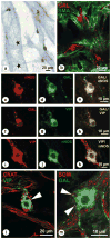
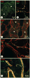
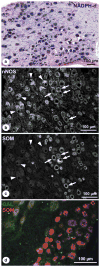
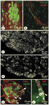
Intrinsic choroidal neurons (ICNs) exist in some primates and bird species. They may act on both vascular and non-vascular smooth muscle cells, potentially influencing choroidal blood flow. Here, we report on the chemical coding of ICNs and eye-related cranial ganglia in the chicken, an important model in myopia research, and further to determine synaptic input onto ICN. Chicken choroid, ciliary, superior cervical, pterygopalatine, and trigeminal ganglia were prepared for double or triple immunohistochemistry of calcitonin gene-related peptide (CGRP), choline acetyltransferase (ChAT), dopamine-beta-hydroxylase, galanin (GAL), neuronal nitric oxide synthase (nNOS), somatostatin (SOM), tyrosine hydroxylase (TH), vasoactive intestinal polypeptide (VIP), vesicular monoamine-transporter 2 (VMAT2), and alpha-smooth muscle actin. For documentation, light, fluorescence, and confocal laser scanning microscopy were used. Chicken ICNs express nNOS/VIP/GAL and do not express ChAT and SOM. ICNs are approached by TH/VMAT2-, CGRP-, and ChAT-positive nerve fibers. About 50% of the pterygopalatine ganglion neurons and about 9% of the superior cervical ganglion neurons share the same chemical code as ICN. SOM-positive neurons in the ciliary ganglion are GAL/NOS negative. CGRP-positive neurons in the trigeminal ganglion lack GAL/SOM. The neurochemical phenotype and synaptic input of ICNs in chicken resemble that of other bird and primate species. Because ICNs lack cholinergic markers, they cannot be readily incorporated into current concepts of the autonomic nervous system. The data obtained provide the basis for the interpretation of future functional experiments to clarify the role of these cells in achieving ocular homeostasis.
Figures


Similar articles
-
Intrinsic neurons in the duck choroid are contacted by CGRP-immunoreactive nerve fibres: evidence for a local pre-central reflex arc in the eye.Exp Eye Res. 2001 Feb;72(2):137-46. doi: 10.1006/exer.2000.0940. Exp Eye Res. 2001. PMID: 11161729
-
Intrinsic choroidal neurons in the duck eye receive sympathetic input: anatomical evidence for adrenergic modulation of nitrergic functions in the choroid.Cell Tissue Res. 2001 May;304(2):175-84. doi: 10.1007/s004410100362. Cell Tissue Res. 2001. PMID: 11396712
-
Intrinsic choroidal neurons in the human eye: projections, targets, and basic electrophysiological data.Invest Ophthalmol Vis Sci. 2003 Sep;44(9):3705-12. doi: 10.1167/iovs.03-0232. Invest Ophthalmol Vis Sci. 2003. PMID: 12939283
-
Choroidal innervation in primate eyes.Exp Eye Res. 2006 Mar;82(3):357-61. doi: 10.1016/j.exer.2005.09.015. Epub 2005 Nov 10. Exp Eye Res. 2006. PMID: 16289045 Review.
-
Innervation of the carotid body: Immunohistochemical, denervation, and retrograde tracing studies.Microsc Res Tech. 2002 Nov 1;59(3):188-95. doi: 10.1002/jemt.10193. Microsc Res Tech. 2002. PMID: 12384963 Review.
Cited by
-
Choroidal and Retinal Abnormalities by Optical Coherence Tomography in Endogenous Cushing's Syndrome.Front Endocrinol (Lausanne). 2016 Dec 9;7:154. doi: 10.3389/fendo.2016.00154. eCollection 2016. Front Endocrinol (Lausanne). 2016. PMID: 28018289 Free PMC article.
-
IMI - Report on Experimental Models of Emmetropization and Myopia.Invest Ophthalmol Vis Sci. 2019 Feb 28;60(3):M31-M88. doi: 10.1167/iovs.18-25967. Invest Ophthalmol Vis Sci. 2019. PMID: 30817827 Free PMC article. Review.
-
Entry of herpes simplex virus type 1 (HSV-1) into the distal axons of trigeminal neurons favors the onset of nonproductive, silent infection.PLoS Pathog. 2012;8(5):e1002679. doi: 10.1371/journal.ppat.1002679. Epub 2012 May 10. PLoS Pathog. 2012. PMID: 22589716 Free PMC article.
-
The choroid-sclera interface: An ultrastructural study.Heliyon. 2022 May 10;8(5):e09408. doi: 10.1016/j.heliyon.2022.e09408. eCollection 2022 May. Heliyon. 2022. PMID: 35586330 Free PMC article.
-
Pharmaceutical intervention for myopia control.Expert Rev Ophthalmol. 2010 Dec 1;5(6):759-787. doi: 10.1586/eop.10.67. Expert Rev Ophthalmol. 2010. PMID: 21258611 Free PMC article.
References
-
- Ahren B, Bottcher G, Kowalyk S, Dunning BE, Sundler F, Taborsky GJ., Jr Galanin is co-localized with noradrenaline and neuropeptide Y in dog pancreas and celiac ganglion. Cell Tissue Res. 1990;261:49–58. - PubMed
-
- Bergua A, Junemann A, Naumann GO. NADPH-D reactive choroid ganglion cells in the human. Klin Monatsbl Augenheilkd. 1993;203:77–82. - PubMed
-
- Bergua A, Mayer B, Neuhuber WL. Nitrergic and VIPergic neurons in the choroid and ciliary ganglion of the duck Anis carina. Anat Embryol. 1996;193:239–248. - PubMed
-
- Brehmer A, Stach W, Krammer HJ, Neuhuber W. Distribution, morphology and projections of nitrergic and non-nitrergic submucosal neurons in the pig small intestine. Histochem Cell Biol. 1998;109:87–94. - PubMed
Publication types
MeSH terms
Grants and funding
LinkOut - more resources
Full Text Sources
Medical
Research Materials

