

Case Reports
doi: 10.1007/s12350-010-9265-8.
Coronary steal: revealing the diagnosis with quantitative cardiac PET/CT
Affiliations
- PMID: 20607629
- PMCID: PMC2990011
- DOI: 10.1007/s12350-010-9265-8
Item in Clipboard
Case Reports
Coronary steal: revealing the diagnosis with quantitative cardiac PET/CT
J Nucl Cardiol.
2010 Dec.
No abstract available
Figures
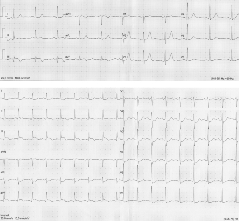
Upper row: baseline ECG compatible with subacute inferior wall myocardial infarction. Lower row: ST-segment elevation in lead III, AVF with reciprocal depression in leads AVL and V2-4 during stress
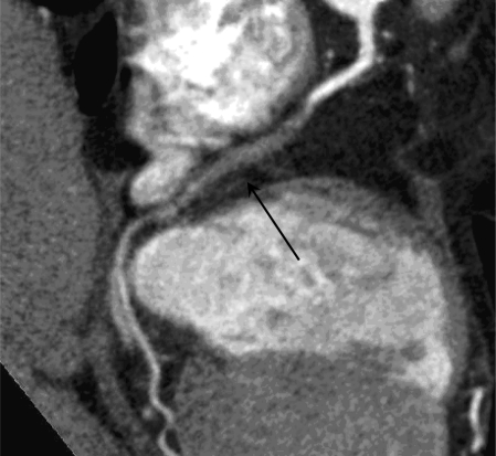
CTCA shows total occlusion of midtraject of the RCA, the distal segment shows contrast opacification caused by collateral filling
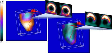
3D fusion of the parametric perfusion PET, with the RCA derived from the CTCA, during rest (left) and vasodilation (right) with corresponding colorscale of absolute perfusion ranging from 0 to 1.0 expressed in mL/minute/g of perfusable tissue. The PET/CT stress images reveal a small perfusion defect during rest which expands to the inferior, inferoseptal, and inferolateral wall during vasodilation
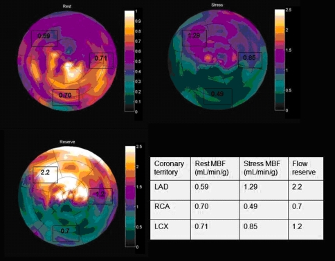
Parametric polar map of absolute quantitative myocardial perfusion (mL/minute/g of perfusable tissue) at rest (right upper quadrant), stress (left upper quadrant), and flow reserve (left lower quadrant). A decreased flow reserve is displayed in inferior, inferolateral, and inferoseptal wall with according quantitative parameters
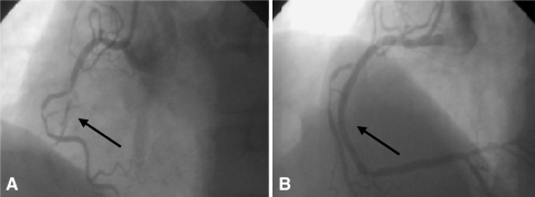
Angiographic images showing the coronary lesion of the RCA before (A) and after stenting (B). Arrow indicates coronary lesion
Similar articles
-
Cardiac PET-CT.J Thorac Imaging. 2007 Feb;22(1):101-6. doi: 10.1097/RTI.0b013e3180317a83. J Thorac Imaging. 2007. PMID: 17325581 Review.
-
A patient with angina (2009: 4a).Eur Radiol. 2009 Apr;19(4):1047. doi: 10.1007/s00330-008-1147-y. Eur Radiol. 2009. PMID: 19277679 No abstract available.
-
Understanding the heart: CT and MRI for coronary heart disease.J Thorac Imaging. 2007 Feb;22(1):107-13. doi: 10.1097/RTI.0b013e3180317457. J Thorac Imaging. 2007. PMID: 17325582 Review.
-
Semi-quantitative myocardial perfusion measured by computed tomography in patients with refractory angina: a head-to-head comparison with quantitative rubidium-82 positron emission tomography as reference.Clin Physiol Funct Imaging. 2017 Sep;37(5):481-488. doi: 10.1111/cpf.12322. Epub 2015 Dec 2. Clin Physiol Funct Imaging. 2017. PMID: 26625937
-
"Mass-ive" infarction: case report and review of myocardial metastatic malignancies.J Nucl Cardiol. 2008 Sep-Oct;15(5):719-26. doi: 10.1016/j.nuclcard.2008.06.017. Epub 2008 Jul 26. J Nucl Cardiol. 2008. PMID: 18761275 Review.
References
-
- Levin DC. Pathways and functional significance of the coronary collateral circulation. Circulation. 1974;50:831–837. - PubMed
-
- Becker L. Conditions for vasodilator-induced coronary steal in experimental myocardial ischemia. Circulation. 1978;57:1103–1110. - PubMed
-
- Patterson R, Kirk E. Coronary steal mechanism in dogs with one-vessel occlusion and other arteries normal. Circulation. 1983;67:1009–1015. - PubMed
-
- Werner GS, Fritzenwanger M, Prochnau D, Schwarz G, Ferrari M, Aarnoudse W, Pijls NHJ, Figulla HR. Determinants of coronary steal in chronic total coronary occlusions: Donor artery, collateral, and microvascular resistance. J Am Coll Cardiol. 2006;48:51–58. doi: 10.1016/j.jacc.2005.11.093. - DOI - PubMed
-
- Seiler C, Fleisch M, Meier B. Direct intracoronary evidence of collateral steal in humans. Circulation. 1997;96:4261–4267. - PubMed
