

Variations in the shape of the frontobasal brain region in obsessive-compulsive disorder
- PMID: 20607751
- PMCID: PMC6869927
- DOI: 10.1002/hbm.21094
Variations in the shape of the frontobasal brain region in obsessive-compulsive disorder
Abstract
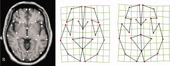
Obsessive-compulsive disorder (OCD) emerges during childhood through young adulthood coinciding with the late phases of postnatal brain development when fine remodeling of brain anatomy takes place. Previous research has suggested the existence of subtle anatomical alterations in OCD involving focal volume variations in different brain regions including the frontal lobes and basal ganglia. We investigated whether anatomical changes might also involve variations in the shape of the frontobasal region. A total of 101 OCD patients and 101 control subjects were examined using magnetic resonance imaging. A cross-sectional image highly representative of frontal-basal ganglia anatomy was selected in each individual and 25 reliable anatomical landmarks were identified to assess shape changes. A pixel-wise morphing approach was also used to dynamically illustrate the findings. We found significant group differences for overall landmark position and for most individual landmarks delimiting the defined frontobasal region. OCD patients showed a deformation pattern involving shortening of the anterior-posterior dimension of the frontal lobes and basal ganglia, and enlargement of cerebrospinal fluid spaces around the frontal opercula. In addition, we observed significant correlation of brain tissue shape variation with frontal sinus size. Identification of a global change in the shape of the frontobasal region may further contribute to characterizing the nature of brain alterations in OCD. The coincidence of brain shape variations with morphological changes in the frontal sinus indicates a potential association of OCD to late development disturbances, as the frontal sinus macroscopically emerges during the transition between childhood and adulthood.
Copyright © 2010 Wiley-Liss, Inc.
Figures
References
-
- Bossa M, Hernandez M, Olmos S ( 2007): Contributions to 3D diffeomorphic atlas estimation: Application to brain images. Med Image Comput Comput Assist Interv 10: 667–674. - PubMed
-
- Brown WA, Molleson TI, Chinn S ( 1984): Enlargement of the frontal sinus. Ann Hum Biol 11: 221–226. - PubMed
-
- Bruner E ( 2007): Cranial shape and size variation in human evolution: Structural and functional perspectives. Childs Nerv Syst 23: 1357–1365. - PubMed
-
- Cardoner N, Soriano‐Mas C, Pujol J, Alonso P, Harrison BJ, Deus J, Hernández‐Ribas R, Menchón JM, Vallejo J ( 2007): Brain structural correlates of depressive comorbidity in obsessive‐compulsive disorder. Neuroimage 38: 413–421. - PubMed
Publication types
MeSH terms
LinkOut - more resources
Full Text Sources
Medical
