. 2010 Nov;225(3):855-64.
doi: 10.1002/jcp.22296.

Agents that bind annexin A2 suppress ocular neovascularization

Affiliations

Agents that bind annexin A2 suppress ocular neovascularization

Raquel Lima e Silva et al. J Cell Physiol. 2010 Nov.

Abstract

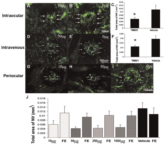
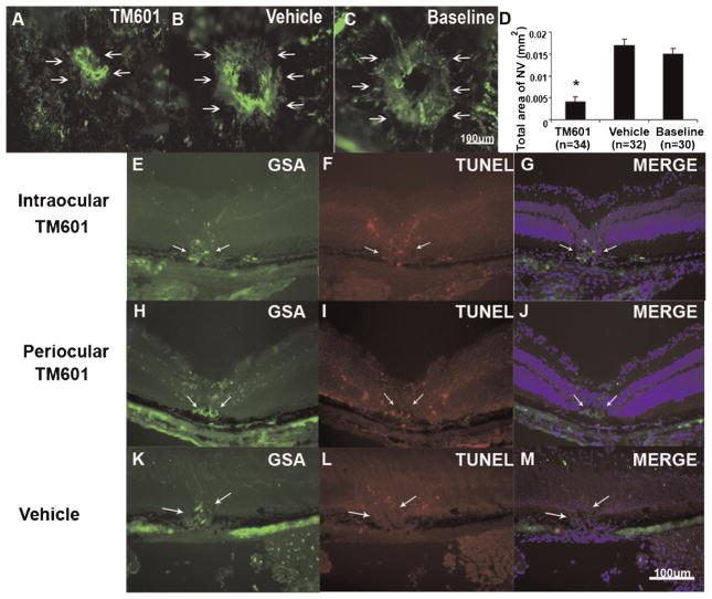
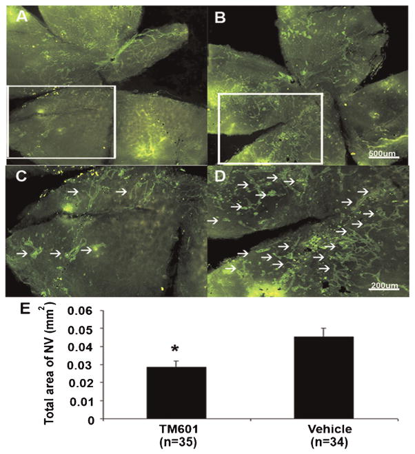
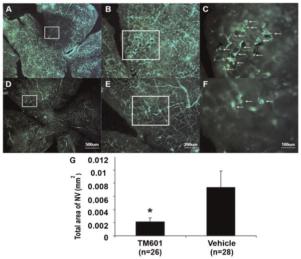
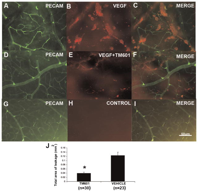
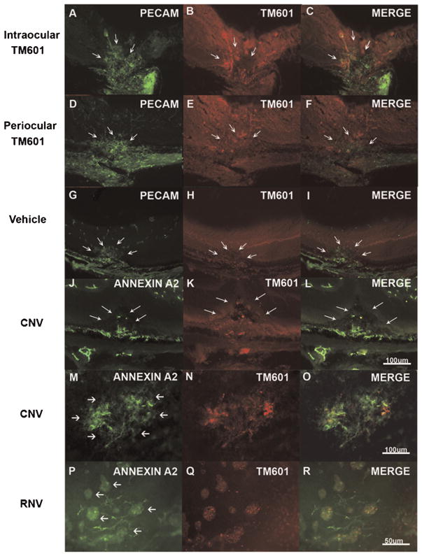
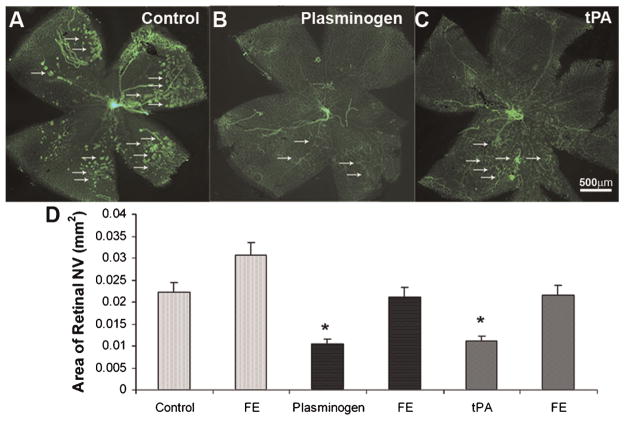
TM601 is a synthetic polypeptide with sequence derived from the venom of the scorpion Leiurus quinquestriatus that has anti-neoplastic activity. It has recently been demonstrated to bind annexin A2 on cultured tumor and vascular endothelial cells and to suppress blood vessel growth on chick chorioallantoic membrane. In this study, we investigated the effects of TM601 in models of ocular neovascularization (NV). When administered by intraocular injection, intravenous injections, or periocular injections, TM601 significantly suppressed the development of choroidal NV at rupture sites in Bruch's membrane. Treatment of established choroidal NV with TM601 caused apoptosis of endothelial cells and regression of the NV. TM601 suppressed ischemia-induced and vascular endothelial growth factor-induced retinal NV and reduced excess vascular permeability induced by vascular endothelial growth factor. Immunostaining with an antibody directed against TM601 showed that after intraocular or periocular injection, TM601 selectively bound to choroidal or retinal NV and co-localized with annexin A2, which is undetectable in normal retinal and choroidal vessels, but is upregulated in endothelial cells participating in choroidal or retinal NV. Intraocular injection of plasminogen or tissue plasminogen activator, which like TM601 bind to annexin A2, also suppressed retinal NV. This study supports the hypothesis that annexin A2 is an important target for treatment of neovascular diseases and suggests that TM601, through its interaction with annexin A2, causes suppression and regression of ocular NV and reduces vascular leakage and thus may provide a new treatment for blinding diseases such as neovascular age-related macular degeneration and diabetic retinopathy.


Figures

Fig. 1
Fig. 1
TM601 given by intraocular, intravenous, or periocular injections suppresses choroidal neovascularization (NV). Mice had laser photocoagulation-induced rupture of Bruch’s membrane and then received an intraocular injection of 1 μl of vehicle or vehicle containing 50 μg of TM601. After 14 days, mice were perfused with fluorescein-labeled dextran and the area of choroidal NV at Bruch’s membrane rupture sites was visualized by fluorescence microscopy of choroidal flat mounts and measured by image analysis. Other groups of mice had rupture of Bruch’s membrane followed by tail vein injections of vehicle or 20 mg/kg of TM601 three times a week. The area of choroidal NV appeared smaller in eyes injected with TM601 (A) compared to those injected with vehicle (B). Statistical comparison using a mixed-effects model showed that the mean (±SEM) area of choroidal NV at Bruch’s membrane rupture sites determined by image analysis with the investigator masked with respect to treatment group was less in eyes injected with TM601 compared to those injected with vehicle (C, P = 0.013). The area of choroidal NV was also significantly reduced in mice that received intravenous injections of TM601 compared to mice that received intravenous injections of vehicle (D–F, P = 0.024). Several different doses of TM601 were given by daily periocular injection. Representative images from eyes injected with 10 μg (G) or 50 μg (H) of TM601 show smaller areas of choroidal NV than a representative image from vehicle-injected eyes (I). Compared to eyes treated with vehicle, the mean (±SEM) area of choroidal NV at Bruch’s membrane rupture sites was significantly less in eyes treated with daily periocular injections of each of the doses of TM601 (J, P = 0.008, 0.001, 0.001, and 0.002 for 10, 50, 250, and 1,000 μg, respectively). Eyes treated with 50 μg (P = 0.007), 250 μg (P = 0.03), or 1,000 μg (P < 0.001) ofTM601 had significantly smaller areas of choroidal NV than their corresponding untreated fellow eyes (FE). There was no significant difference in area of choroidal NV in eyes treated with 10 μg ofTM601 compared to corresponding fellow eyes (P = 0.147).
Fig. 2
Fig. 2
Intraocular injection of TM601 causes regression of established choroidal neovascularization (NV). Seven days after rupture of Bruch’s membrane with laser photocoagulation, the baseline area of choroidal NV was measured in 10 mice. The 12 remaining mice were given an intraocular injection of 1 μl of vehicle in one eye and 1 μl of vehicle containing 50 μg of TM601 in the other eye. Seven days later, the area of choroidal NV at rupture sites appeared smaller in eyes injected with TM601 (A) compared to those in vehicle-injected eyes (B) or baseline eyes (C). Measurement of the area of choroidal NV by image analysis (D) showed that the mean area of choroidal NV in eyes injected with TM601 was significantly less than that in vehicle-injected or those in baseline eyes (P = 0.002 and 0.018 by a general estimating equations model). Mice had rupture of Bruch’s membrane and after 1 week had an intraocular injection of 50 μg of TM601 in one eye (E–G) and vehicle in the fellow eye (K–M) or had daily periocular injections of 250 μg of TM601 (H–J). After 2 days, ocular sections through choroidal NV showed staining of the NV with Griffonia simplicifolia lectin (GSA), a vascular endothelial cell marker (E,H,K, arrows). Sections from eyes treated with TM601 showed TUNEL-positive, GSA-positive endothelial cells in the choroidal NV and also in the overlying retina from the laser treatment that induced the choroidal NV (F,G,I,J). Sections from vehicle-treated eyes showed TUNEL-positive cells in overlying retina, but none in the GSA-stained choroidal NV (L,M).
Fig. 3
Fig. 3
TM601 suppresses ischemia-induced retinal neovascularization. At postnatal day (P) 7, mice were placed in 75% oxygen and at P12 they were returned to room air and given an intraocular injection of 1 μl containing 50 μg of TM601 in one eye and vehicle in the other eye. At P17, in vivo immunostaining for PECAM-1 was done and retinal whole mounts were examined by fluorescence microscopy. Low power views of entire retinas from representative TM601-treated (A) and vehicle-treated (B) eyes show less neovascularization on the surface of the TM601-treated retina. Higher magnification of the boxed regions from (A) and (B) provide better resolution of individual tufts of neovascularization that are reduced in TM601-treated eyes (C) compared to vehicle-treated eyes (D). Image analysis with the investigator masked with respect to treatment group, showed that the mean (±SEM) area of NV on the surface of the retina was significantly less in TM601-treated eyes compared to those treated with vehicle (E, P = 0.006 by mixed-effects model).
Fig. 4
Fig. 4
TM601 suppresses VEGF-induced subretinal neovascularization (NV) in rhodopsin promoter/VEGF (rho/VEGF) transgenic mice. Between P7 and P21 rho/VEGF mice were given daily periocular injections of 3 μl of vehicle or vehicle containing 150 μg of TM601. Mice were perfused with fluorescein-labeled dextran at P21. Retinal flat mounts from control, vehicle-treated eyes visualized with the photoreceptor side facing up showed extensive NV on the outer surface of the retina although it is difficult to see at the magnification that allows visualization of the entire retina because the normal retinal vessels are in the background (A). Higher magnification views (B,C) of the boxed region in (A) provides an arrow depth of field so that only the NV is in focus and the numerous tufts of NV that are partially surrounded by dark retinal pigmented epithelial cells are easily seen (C, arrows). Retinal flat mounts from TM601-injected eyes show much less NV (D); higher magnification of the boxed region in (D) shows few tufts of NV on the outer surface of the retina (E,F, arrows). Image analysis with the investigator masked with respect to treatment group showed that the mean (±SEM) area of NV per retina was significantly less fore yes treated with TM601 compared to controls (G). *P = 0.017 for difference from control by mixed-effects model.
Fig. 5
Fig. 5
TM601 suppresses VEGF-induced retinal vascular permeability. Six hours after intraocular injections of VEGF, a mixture of VEGF and TM601, or vehicle, mice were euthanized and retinas were stained for serum albumin or PECAM-1. Retinas from eyes injected with vehicle only, showed no staining for albumin (G–I), when examined by fluorescence microscopy. Retinas from eyes injected with VEGF alone showed significantly more large aggregates of albumin adjacent to retinal vessels (A–C) comparing to retinas from eyes injected with a mixture of VEGF and TM601 (D–F). Measurement of the leakage area of albumin showed that TM601 significantly reduced VEGF-induced leakage of albumin into the retina (P = 0.0001 by ANOVA with Bonferroni/Dunn test).
Fig. 6
Fig. 6
TM601 homes to retinal and choroidal neovascularization (NV) where it is co-localized with upregulated annexin A2. Mice had rupture of Bruch’s membrane and after 1 week had an intraocular injection of 50 μg of TM601 in one eye (A–C) and vehicle in the fellow eye (G–I) or had daily periocular injections of 250 μg of TM601 (D–F). After 2 days, ocular sections through choroidal NV showed staining for PECAM-1 in the NV (A, arrows) and underlying choroidal vessels. Staining of the same section with an antibody that specifically recognizes TM601 showed strong immunofluorescence with a shape similar to the anti-PECAM-1 staining (B, arrows) and some mild background fluorescence throughout the retina. Merging of the images showed clear co-localization of TM601 and PECAM-1 (C, arrows). Eyes given periocular injections of TM601 also showed strong staining for TM601 in PECAM-1-labeled choroidal NV (D–F, arrows). Eyes given an intraocular injection of vehicle showed PECAM-1 staining of choroidal NV (G, arrows), but no staining for TM601 (H,I, arrows). Sections from eyes injected with TM601 showed staining for annexin A2 in choroidal NV (CNV,J, arrows) that co-localized with staining for TM601 (K,L, arrows). Achoroidal flat mount from an eye that had intraocular injection of TM601 7 days after rupture of Bruch’s membrane showed intense staining of choroidal NV with anti-annexin A2 with no staining of surrounding choroidal vessels (M, arrows). There was also strong staining for TM601 thatco-localized with the staining for annexin A2 and with this technique there was minimal background staining (N,O). Similar experiments were done in mice with ischemic retinopathy to determine if annexin A2 is also upregulated in retinal NV and whether TM601 binds to retinal NV. At P17, mice with ischemic retinopathy were given and intraocular injection of 50 μg of TM601 and at P19, retinal flat mounts showed strong selective staining for annexin A2 on tufts of retinal NV (RNV; P, arrows). Punctate staining for TM601 also occurred within the retinal NV (Q,R) indicating that annexin A2 is upregulated in retinal NV and TM601 binds in the areas of annexin A2 upregulation.
Fig. 7
Fig. 7
Effects of tPA or plasminogen in ischemia-induced retinal neovascularization (NV). C57BL/6 mice were placed in 75 ± 3% oxygen at P7. At P12 they were returned to room air and were given an intravitreous injection of 1 μl of vehicle or vehicle containing 0.5 μg of plasminogen or tissue plasminogen activator (tPA) in one eye. At P17, in vivo immunostaining for PECAM-1 was done and retinal flat mounts from eyes injected with plasminogen (B) or tPA (C) visualized with fluorescence microscopy showed less NV on the surface of the retina than eyes injected with vehicle (A). Quantification of the area of NV by image analysis is shown in (D). The bars show the mean (±SEM) area of NV and those for plasminogen (n =13) and tPA (n =8) were significantly less than respective control eyes injected with vehicle (P < 0.05 by unpaired t-test) or uninjected fellow eyes (FE).

References

    1. Brown DM, Kaiser PK, Michels M, Soubrane G, Heier JS, Kim RY, Sy JP, Schneider S Anchor Study Group. Ranibizumab versus verteporfin for neovascular age-related macular degeneration. N Engl J Med. 2006;355:1432–1444. - PubMed
    1. Campochiaro PA. Ocular versus extraocular neovascularization: Mirror images or vague resemblances. Invest Ophthalmol Vis Sci. 2006;47:462–474. - PubMed
    1. Cesarman GM, Guevara CA, Hajjar KA. An endothelial cell receptor for plasminogen and tissue plasminogen activator. II. Annexin II-mediated enhancement of t-PA-dependent plasminogen activation. J Biol Chem. 1994;269:21198–21203. - PubMed
    1. DeBin JA, Maggio JE, Strichartz GR. Purification and characterization of chlorotoxin, a chloride channel ligand from the venom of the scorpion. Am J Physiol. 1993;264:C361–C369. - PubMed
    1. Deora AB, Kreitzer G, Jacovina AT, Hajjar KA. An annexin 2 phosphorylation switch mediates p11-dependent translocation of annexin 2 to the cell surface. J Biol Chem. 2004;279:43411–43418. - PubMed

Publication types

MeSH terms

LinkOut - more resources