

Cisplatin and PI3kinase inhibition decrease invasion and migration of human ovarian carcinoma cells and regulate matrix-metalloproteinase expression
- PMID: 20607860
- PMCID: PMC3001291
- DOI: 10.1002/cm.20465
Cisplatin and PI3kinase inhibition decrease invasion and migration of human ovarian carcinoma cells and regulate matrix-metalloproteinase expression
Abstract
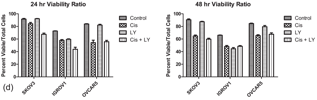
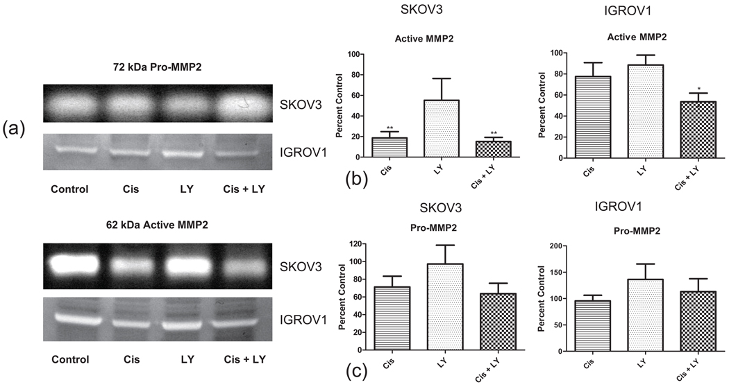
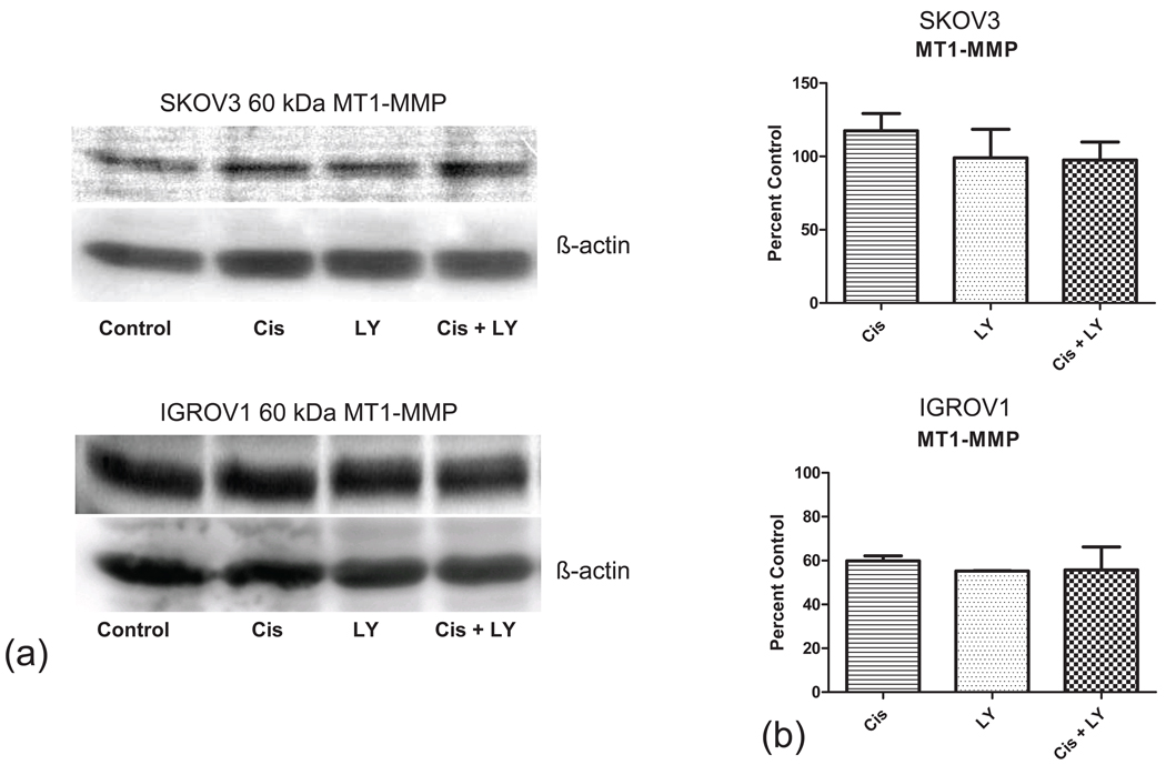
Targeting of the PI3K (phosphoinositide3-kinase)/Akt/mTOR pathway in human ovarian cancer cells is a promising novel therapeutic strategy. We investigated the effects of cisplatin and the PI3K inhibitor LY294002 on invasion, migration and the expression of essential matrix metalloproteinases (MMPs) in ovarian cancer cells. SKOV3, OVCAR5 and IGROV1 human ovarian cancer cell lines were treated with cisplatin, LY294002 and a combination of both drugs. Invasion and migration of treated cells was assessed using Matrigel and uncoated PET membrane assays. Expression levels of pro-MMP2, MMP2, TIMP1, TIMP2 and MT1-MMP were determined using Western Blotting. Gel zymography was used to quantitate the functional levels of active MMP2. All three cell lines showed significantly reduced invasion and migration after treatment with cisplatin, LY294002, and the combination of both drugs compared to untreated controls. In SKOV3 cells, cisplatin alone and in combination with LY294002 resulted in a 6.3 and 7.1-fold reduction in the total amount of activated MMP2. TIMP1 expression decreased by 5.0, 6.6 and 28.4-fold with cisplatin, LY294002 and the combination respectively (P < 0.05). In contrast, only cisplatin and the combination of both drugs resulted in a significant, 3.7 and 5.1-fold reduction in the level of TIMP2. Expression levels of MT1-MMP remained unchanged. These observations were corroborated in IGROV1 cell lines that showed similar changes of activated MMP2 and TIMP2 expression, but no significant decrease in TIMP1 levels. Our data suggests that inhibition of ovarian cancer cell motility is mediated via down-regulation of activated MMP2, TIMP1 and TIMP2 expression under these treatment conditions.
2010 Wiley-Liss, Inc.
Figures


Similar articles
-
Dual targeting of phosphoinositide 3-kinase and mammalian target of rapamycin using NVP-BEZ235 as a novel therapeutic approach in human ovarian carcinoma.Clin Cancer Res. 2011 Apr 15;17(8):2373-84. doi: 10.1158/1078-0432.CCR-10-2289. Epub 2011 Mar 3. Clin Cancer Res. 2011. PMID: 21372221 Free PMC article.
-
[Combined inhibition of PI3K and MEK has synergistic inhibitory effect on the proliferation of cisplatin-resistant ovarian cancer cells].Xi Bao Yu Fen Zi Mian Yi Xue Za Zhi. 2014 Jun;30(6):592-6. Xi Bao Yu Fen Zi Mian Yi Xue Za Zhi. 2014. PMID: 24909280 Chinese.
-
HPIP promotes epithelial-mesenchymal transition and cisplatin resistance in ovarian cancer cells through PI3K/AKT pathway activation.Cell Oncol (Dordr). 2017 Apr;40(2):133-144. doi: 10.1007/s13402-016-0308-2. Epub 2016 Dec 30. Cell Oncol (Dordr). 2017. PMID: 28039608
-
AKT and mTOR phosphorylation is frequently detected in ovarian cancer and can be targeted to disrupt ovarian tumor cell growth.Oncogene. 2004 Jul 29;23(34):5853-7. doi: 10.1038/sj.onc.1207721. Oncogene. 2004. PMID: 15208673
-
Targeting the PI3K pathway and DNA damage response as a therapeutic strategy in ovarian cancer.Cancer Treat Rev. 2020 Jun;86:102021. doi: 10.1016/j.ctrv.2020.102021. Epub 2020 Apr 10. Cancer Treat Rev. 2020. PMID: 32311593 Free PMC article. Review.
Cited by
-
Minocycline attenuates hypoxia-inducible factor-1α expression correlated with modulation of p53 and AKT/mTOR/p70S6K/4E-BP1 pathway in ovarian cancer: in vitro and in vivo studies.Am J Cancer Res. 2015 Jan 15;5(2):575-88. eCollection 2015. Am J Cancer Res. 2015. PMID: 25973298 Free PMC article.
-
MicroRNA-509-3p inhibits cellular migration, invasion, and proliferation, and sensitizes osteosarcoma to cisplatin.Sci Rep. 2019 Dec 13;9(1):19089. doi: 10.1038/s41598-019-55170-2. Sci Rep. 2019. PMID: 31836741 Free PMC article.
-
Regulation of ATP utilization during metastatic cell migration by collagen architecture.Mol Biol Cell. 2018 Jan 1;29(1):1-9. doi: 10.1091/mbc.E17-01-0041. Epub 2017 Nov 8. Mol Biol Cell. 2018. PMID: 29118073 Free PMC article.
-
Dual targeting of phosphoinositide 3-kinase and mammalian target of rapamycin using NVP-BEZ235 as a novel therapeutic approach in human ovarian carcinoma.Clin Cancer Res. 2011 Apr 15;17(8):2373-84. doi: 10.1158/1078-0432.CCR-10-2289. Epub 2011 Mar 3. Clin Cancer Res. 2011. PMID: 21372221 Free PMC article.
-
Inhibition of Phosphatidylinositol 3-kinase (PI3K) Signaling Synergistically Potentiates Antitumor Efficacy of Paclitaxel and Overcomes Paclitaxel-Mediated Resistance in Cervical Cancer.Int J Mol Sci. 2019 Jul 10;20(14):3383. doi: 10.3390/ijms20143383. Int J Mol Sci. 2019. PMID: 31295843 Free PMC article.
References
-
- Altomare DA, Testa JR. Perturbations of the AKT signaling pathway in human cancer. Oncogene. 2005;24(50):7455–7464. - PubMed
-
- Altomare DA, Wang HQ, Skele KL, De Rienzo A, Klein-Szanto AJ, Godwin AK, Testa JR. AKT and mTOR phosphorylation is frequently detected in ovarian cancer and can be targeted to disrupt ovarian tumor cell growth. Oncogene. 2004;23(34):5853–5857. - PubMed
-
- Atkinson SJ, Crabbe T, Cowell S, Ward RV, Butler MJ, Sato H, Seiki M, Reynolds JJ, Murphy G. Intermolecular autolytic cleavage can contribute to the activation of progelatinase A by cell membranes. J Biol Chem. 1995;270(51):30479–30485. - PubMed
-
- Bellacosa A, Kumar CC, Di Cristofano A, Testa JR. Activation of AKT kinases in cancer: implications for therapeutic targeting. Adv Cancer Res. 2005;94:29–86. - PubMed
-
- Bjorklund M, Koivunen E. Gelatinase-mediated migration and invasion of cancer cells. Biochim Biophys Acta. 2005;1755(1):37–69. - PubMed
Publication types
MeSH terms
Substances
Grants and funding
LinkOut - more resources
Full Text Sources
Other Literature Sources
Medical
Research Materials
Miscellaneous
