Comparative Study
. 2010 Oct;51(10):2011-22.
doi: 10.1111/j.1528-1167.2010.02652.x.

Simultaneous EEG, fMRI, and behavior in typical childhood absence seizures

Affiliations
Comparative Study

Simultaneous EEG, fMRI, and behavior in typical childhood absence seizures

Rachel Berman et al. Epilepsia. 2010 Oct.

Abstract

Purpose: Absence seizures cause transient impairment of consciousness. Typical absence seizures occur in children, and are accompanied by 3-4-Hz spike-wave discharges (SWDs) on electroencephalography (EEG). Prior EEG-functional magnetic resonance imaging (fMRI) studies of SWDs have shown a network of cortical and subcortical changes during these electrical events. However, fMRI during typical childhood absence seizures with confirmed impaired consciousness has not been previously investigated.

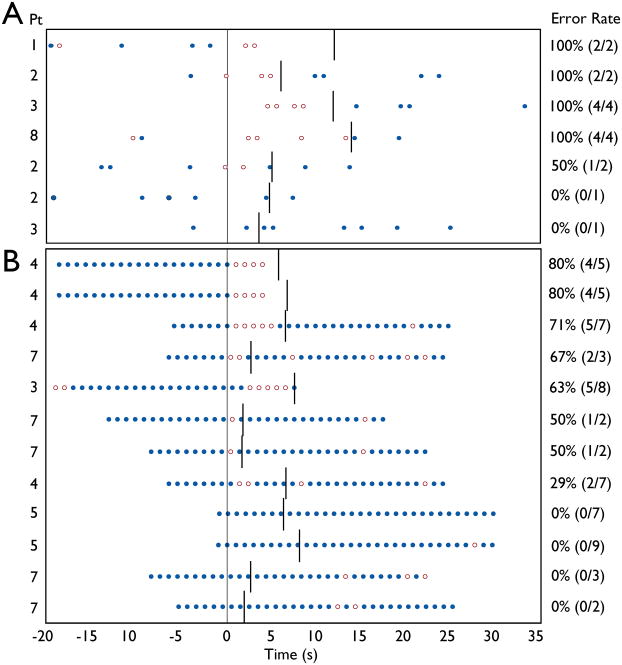
Methods: We performed EEG-fMRI with simultaneous behavioral testing in 37 children with typical childhood absence epilepsy (CAE). Attentional vigilance was evaluated by a continuous performance task (CPT), and simpler motor performance was evaluated by a repetitive tapping task (RTT).

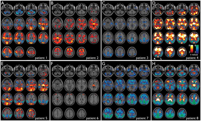
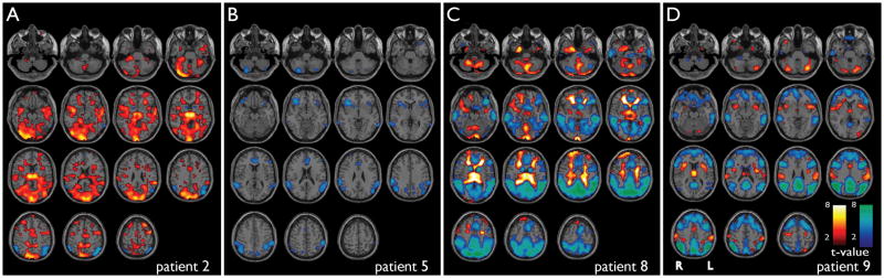
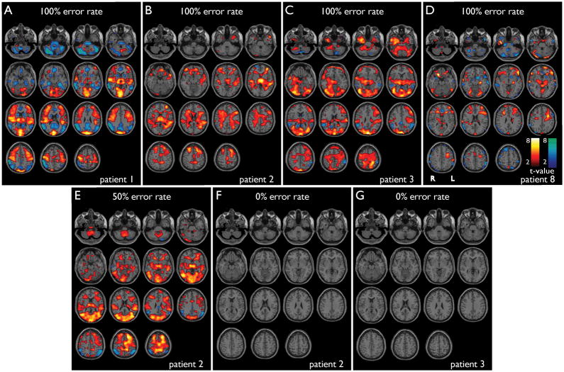
Results: SWD episodes were obtained during fMRI scanning from 9 patients among the 37 studied. fMRI signal increases during SWDs were observed in the thalamus, frontal cortex, primary visual, auditory, somatosensory, and motor cortex, and fMRI decreases were seen in the lateral and medial parietal cortex, cingulate gyrus, and basal ganglia. Omission error rate (missed targets) with SWDs during fMRI was 81% on CPT and 39% on RTT. For those seizure epochs during which CPT performance was impaired, fMRI changes were seen in cortical and subcortical structures typically involved in SWDs, whereas minimal changes were observed for the few epochs during which performance was spared.

Discussion: These findings suggest that typical absence seizures involve a network of cortical-subcortical areas necessary for normal attention and primary information processing. Identification of this network may improve understanding of cognitive impairments in CAE, and may help guide development of new therapies for this disorder.


Conflict of interest statement

None of the authors has any conflict of interest to disclose.

Figures

Figure 1
Figure 1
Cortical and subcortical fMRI changes during absence seizures in 8 patients while undergoing behavioral testing. Six motion-related parameters (3 translation, 3 rotation) are included in the modeling. Axial sections show BOLD fMRI increases (warm colors) and decreases (cool colors) during seizures compared to baseline analyzed in SPM. fMRI increases were seen most consistently in the thalamus, medial occipital, lateral temporal (Heschl’s), superior Rolandic, and lateral frontal cortex, and decreases were seen in lateral parietal, medial parietal, cingulate cortex and basal ganglia. a) Patient 1, seizure duration =12.0 s. b) Patient 2, seven seizures (mean duration = 4.9 s). c) Patient 3, three seizures (mean duration = 7.8 s). d) Patient 4, six seizures (mean duration = 6.0 sec). e) Patient 5, six seizures (mean duration = 6.8 s). f) Patient 6, three seizures (mean duration = 5.8 s). g) Patient 7, seven seizures (mean duration = 2.4 s). h) Patient 8, nine seizures (mean duration = 11.4 s). SPM analysis threshold P=0.05, FDR-corrected, and extent threshold k=3 voxels.
Figure 2
Figure 2
fMRI changes during absence seizures in 4 patients during fixation only (no task) with 6 motion related parameters (3 translation, 3 rotation) included in the modeling. Results were generally similar to fMRI changes in the same patients with seizures during behavioral tasks (Figure 1), showing increases in the thalamus, medial occipital, temporal and lateral frontal cortex, and decreases in the parietal cortex, cingulate gyrus and basal ganglia. Axial sections show BOLD fMRI increases (warm colors) and decreases (cool colors) during seizures compared to baseline analyzed in SPM. a) Patient 2, two seizures (mean duration = 2.3 s). Compare to same patient in Figure 1b. b) Patient 5, one seizure (duration = 7.5 s). Compare to same patient in Figure 1e. c) Patient 8, nine seizures (mean duration = 7.0 s). Compare to same patient in Figure 1h. d) Patient 9, 6 seizures (mean duration = 1.9 s). SPM analysis threshold P=0.05, FDR-corrected, and extent threshold k=3 voxels.
Figure 3
Figure 3
Behavioral performance on CPT and RTT tasks during absence seizures. Behavioral tasks consisted of 32 s blocks of letter presentations, alternating with 32 s of fixation. Letters were presented once per second. a) For CPT, the target (X) occurred at random, at an average rate of once per 4 seconds. Mean omission error rate during seizures was 81%.(13/16). b) During RTT, patients were instructed to push the button for all letters (no Xs were presented). Mean omission error rate during seizures was 39% (24/60). ●= correct response to target. ○ = omission error. Symbols appear at time of letter presentation. Response was required within 1000 ms of letter presentation. Vertical line at t = 0 is seizure onset. Other vertical lines denote seizure end. 22 seizures were recorded during task blocks, but two seizures had no target letters occurring during the spike-wave discharge, and one could not be analyzed due to technical difficulty with button press recording, resulting in 19 seizures total.
Figure 4
Figure 4
fMRI changes in seizures with good vs. poor performance on CPT. SPM results for individual seizures are shown. (a–e) Seizures during impaired performance on CPT, showing cortical and subcortical fMRI changes. (f, g) Seizures during good performance on CPT (omission error rate 0%) show no significant cortical or subcortical changes on fMRI. a) Patient 1, seizure duration =12.0 s, omission error rate = 100%. b) Patient 2, seizure duration = 6.0 s, omission error rate = 100%. c) Patient 3, seizure duration = 11.9 s, omission error rate = 100%. d) Patient 8, seizure duration = 14.0 s, omission error rate = 100%. e) Patient 2, seizure duration = 5.0 s, omission error rate = 50%. f) Patient 2, seizure duration = 4.7 s, omission error rate = 0%. g) Patient 3, seizure duration = 3.8 s, omission error rate = 0%. Performance data on CPT during these same seizures is shown in Figure 3a. Warm colors represent fMRI increases, and cool colors decreases. SPM analysis threshold P=0.05, FDR-corrected, and extent threshold k=3 voxels, with 6 motion-related parameters included in the model.

Similar articles

Cited by

References

    1. Aghakhani Y, Bagshaw AP, Benar CG, Hawco C, Andermann F, Dubeau F, Gotman J. fMRI activation during spike and wave discharges in idiopathic generalized epilepsy. Brain. 2004;127:1127–1144. - PubMed
    1. Archer JS, Abbott DF, Waites AB, Jackson GD. fMRI “deactivation” of the posterior cingulate during generalized spike and wave. Neuroimage. 2003;20:1915–1922. - PubMed
    1. Avoli M, Gloor P, Kostopoulos G, Naquet T, editors. Generalized Epilepsy. Boston: Birkhauser; 1990.
    1. Bai X, Vestal M, Berman R, Negishi M, Spann M, Vega C, DeSalvo MN, Novotny E, Constable RT, Blumenfeld H. Dynamic timecourse of typical childhood absence seizures: EEG, behavior and fMRI. J Neurosci. 2010 In Press. - PMC - PubMed
    1. Blumenfeld H. The thalamus and seizures. Arch Neurol. 2002;59:135–137. - PubMed

Publication types

MeSH terms