. 2010 Oct;36(6):462-77.
doi: 10.1111/j.1365-2990.2010.01103.x.

Tyrosine phosphorylation of tau accompanies disease progression in transgenic mouse models of tauopathy

Affiliations

Tyrosine phosphorylation of tau accompanies disease progression in transgenic mouse models of tauopathy

K Bhaskar et al. Neuropathol Appl Neurobiol. 2010 Oct.

Abstract

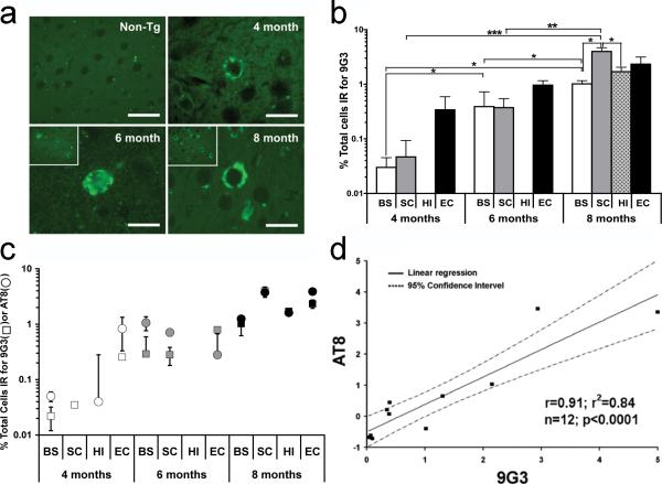
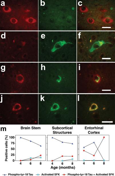
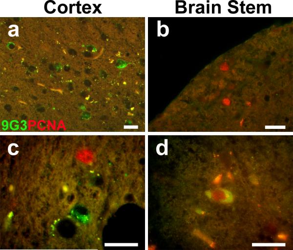
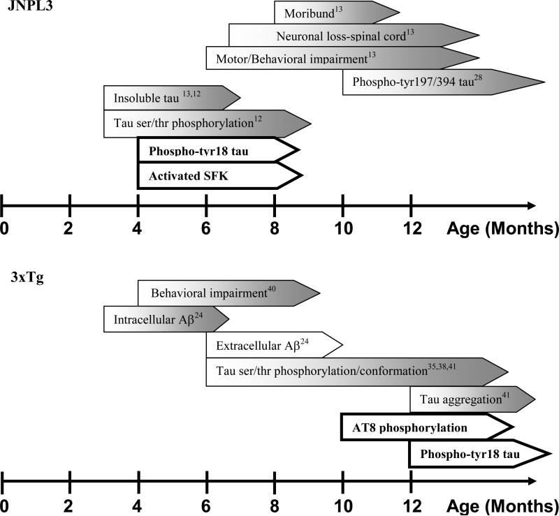
Aim: Tau protein is a prominent component of paired helical filaments in Alzheimer's disease (AD) and other tauopathies. While the abnormal phosphorylation of tau on serine and threonine has been well established in the disease process, its phosphorylation on tyrosine has only recently been described. We previously showed that the Src family non-receptor tyrosine kinases (SFKs) Fyn and Src phosphorylate tau on Tyr18 and that phospho-Tyr18-tau was present in AD brain. In this study, we have investigated the appearance of phospho-Tyr18-tau, activated SFK and proliferating cell nuclear antigen (PCNA) during disease progression in a mouse model of human tauopathy.

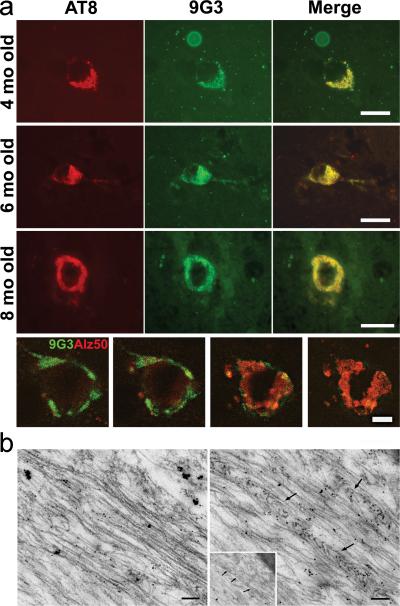
Methods: We have used JNPL3, which expresses human tau with P301L mutation, and antibodies specific for phospho-Tyr18-tau (9G3), ser/thr phosphorylated tau (AT8), activated SFK and PCNA. Antibody staining was viewed by either epifluorescence or confocal microscopy.

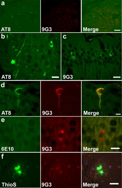
Results: Phospho-Tyr18-tau appeared concurrently with AT8-reactive tau as early as 4 months in JNPL3. Some 9G3-positive cells also contained activated SFKs and PCNA. We also investigated the triple transgenic mouse model of AD and found that unlike the JNPL3 model, the appearance of 9G3 reactivity did not coincide with AT8 in the hippocampus, suggesting that the presence of APP/presenilin influences tau phosphorylation. Also, Thioflavin S-positive plaques were 9G3-negative, suggesting that phospho-Tyr18-tau is absent from the dystrophic neurites of the mouse triple transgenic brain.

Conclusions: Our results provide evidence for the association of tyrosine-phosphorylated tau with mechanisms of neuropathogenesis and indicate that SFK activation and cell cycle activation are also involved in JNPL3.


Figures

Fig. 1
Fig. 1
(a) Paraffin sections from 4, 6, and 8 month JNPL3 mouse brain (entorhinal cortex) were labeled with 9G3 and viewed by epifluorescence. Scale bar: 20μm. Insets at lower magnification show that 8 month old sample displayed increased number of phospho-Tyr18 tau positive cells relative to 6 month old. Phospho-Tyr18 tau immunoreactivity was lacking in 8-month-old non-transgenic mouse (upper left, scale bar: 30μm). (b) Phospho-Tyr18 tau appears at four months in three of the four brain regions and steadily increases with age in JNPL3 mice. Percentage of cells positive for 9G3 (number of positive cells divided by total number of cells examined in each brain region, as determined by Hoechst staining) was calculated from each animal (n=3). Values are expressed in mean percentage ±SEM. Comparisons were made between different brain regions within the same age group and for the same brain regions across different age groups. Differences that were statistically significant by unpaired t-test are indicated (*p<0.05; **p<0.01; ***p<0.005). BS-Brain Stem; SC-SubCortical structures; HI-Hippocampus; EC-Entorhinal Cortex (c) Correlation plot showing that the percentage of cells (mean+SEM) labeled for phospho-Tyr18 tau (squares) or AT8 (circles) increased with age in different brain regions of JNPL3 mice. AT8 or 9G3 positive cells were counted following single labeling. Individual markers (square or circle) within each age group represent the percentage of positive cells in brain stem (BS), subcortical structures (SC), hippocampus (HI) and entorhinal cortex (EC). Total number of cells examined per region (100%) was based on Hoechst staining. (d) Linear regression analysis between percentage of AT8 and 9G3 positive cells showed a significant correlation between 9G3 and AT8 immunoreactive cells in the different brain regions of each age group studied.
Fig. 2
Fig. 2
(a) Representative photomicrographs of the entorhinal cortices of 4-, 6- and 8-months-old JNPL3 mice brain showing cells double labeled with AT8 (red-left panel) and 9G3 (green-middle panel); merged images are shown in right panel. Scale bar: 20 μm. The bottom row shows four optical sections (9, 11, 14, 16) from a z-series stack of 22 optical sections (0.5μm thick) stained for 9G3 (green) and Alz50 (red) in the 8-month-old cortex. 9G3 positive tau was most abundant in sections 9-11 whereas Alz50 immunoreactive tau was most abundant in sections 14-16. Scale bar: 10 μm. (b) Post-embedding immunogold labeling with 9G3 showed 10nm gold particles decorating straight filaments (left) and filaments forming a herringbone pattern (right arrows) in ultrathin sections from JNPL3 spinal cord. Scale bar: 0.1μm.
Fig. 3
Fig. 3
Appearance of phospho-Tyr18 tau in 3xTg mouse brain. (a) Each panel shows a confocal projection generated from z-series stack of 10 optical sections (0.5μm apart) AT8 immunoreactivity was observed in the fimbria region of 10 month old 3xTg brain. 9G3 labeling was absent. Scale bar: 30 μm. (b) AT8 immunoreactive dystrophic neurites were observed in the subiculum of 12 month old 3xTg brain. Scale bar: 20 μm. (c) 9G3 labeling was observed in the hippocampus. Scale bar: 20 μm. (d) Co-localization of AT8 and 9G3 was observed in 12 month old CA1 pyramidal neuron following double labeling with AT8 (green) and biotinylated-9G3 (red). Scale bar: 10 μm. (e) 9G3 positive CA1 hippocampal neurons lacked 6E10 intracellular Aβ staining. Scale bar: 15 μm. (f) Mature amyloid plaque, identified by Thio S, lacked 9G3 staining. Scale bar: 20 μm. (b-f) were viewed by epifluorescence microscopy.
Fig. 4
Fig. 4
Representative photomicrographs from the brain stem of 8-month-old JNPL3 mouse showing cells immunoreactive for only activated SFK (a-c) or phospho-Tyr18 tau (d-f). When activated SFK and phospho-Tyr18 appeared in the same cell in the entorhinal cortex, the subcellular distribution was either different (g-i) or overlapping (jl). Scale bar: 20μm. (m) Percentage of positive cells immunoreactive for phospho-Tyr18 tau (9G3) alone (dark blue triangles), active-Src (pY416) alone (light blue squares) or for both (red circles) with increasing age in different brain regions of JNPL3 mice. The total number of positive cells per age group in respective brain regions was counted following double labeling with 9G3 and pY416 antibodies and assigned 100%. Hippocampus had no labeled cells at 4 and 6 months, then showed only double-labeled cells (both 9G3 and pY416 positive) at 8 months.
Fig. 5
Fig. 5
Partial overlap in the expression pattern of phospho-Tyr18 tau and PCNA in the JNPL3 mouse brain. Representative overlay images from 8-month-old JNPL3 double stained for phospho-tyr 18 (green) and PCNA (red) showed (a) cells immunoreactive for only phospho-Tyr18 tau in cortex, (b) cells immunoreactive for only PCNA in the brainstem, (c) both cell types in a single visual field in the cortex, and (d) cells immunoreactive for both phospho-Tyr18 and PCNA in the brainstem. Scale bar: 20μm.
Fig. 6
Fig. 6
Summary of the relationship between phosphorylation of tau on Tyr18 and other attributes of JNPL3 (top) or 3xTg (bottom). Findings from this study are bolded.

Similar articles

Cited by

References

    1. Goedert M. Tau protein and the neurofibrillary pathology of Alzheimer's disease. Trends Neurosci. 1993;16:460–5. - PubMed
    1. Williamson R, Scales T, Clark BR, Gibb G, Reynolds CH, Kellie S, Bird IN, Varndell IM, Sheppard PW, Everall I, Anderton BH. Rapid tyrosine phosphorylation of neuronal proteins including tau and focal adhesion kinase in response to amyloid-beta peptide exposure: involvement of Src family protein kinases. J Neurosci. 2002;22:10–20. - PMC - PubMed
    1. Derkinderen P, Scales TM, Hanger DP, Leung KY, Byers HL, Ward MA, Lenz C, Price C, Bird IN, Perera T, Kellie S, Williamson R, Noble W, Van Etten RA, Leroy K, Brion JP, Reynolds CH, Anderton BH. Tyrosine 394 is phosphorylated in Alzheimer's paired helical filament tau and in fetal tau with c-Abl as the candidate tyrosine kinase. J Neurosci. 2005;25:6584–93. - PMC - PubMed
    1. Lee G, Thangavel R, Sharma VM, Litersky JM, Bhaskar K, Fang SM, Do LH, Andreadis A, Van Hoesen G, Ksiezak-Reding H. Phosphorylation of tau by fyn: implications for Alzheimer's disease. J Neurosci. 2004;24:2304–12. - PMC - PubMed
    1. Lebouvier T, Scales TM, Hanger DP, Geahlen RL, Lardeux B, Reynolds CH, Anderton BH, Derkinderen P. The microtubule-associated protein tau is phosphorylated by Syk. Biochim Biophys Acta. 2008;1783:188–92. - PMC - PubMed

Publication types