

Tissue microarrays: one size does not fit all
- PMID: 20609235
- PMCID: PMC2910003
- DOI: 10.1186/1746-1596-5-48
Tissue microarrays: one size does not fit all
Abstract
Background: Although tissue microarrays (TMAs) are commonly employed in clinical and basic-science research, there are no guidelines for evaluating the appropriateness of a TMA for a given biomarker and tumor type. Furthermore, TMA performance across multiple biomarkers has not been systematically explored.
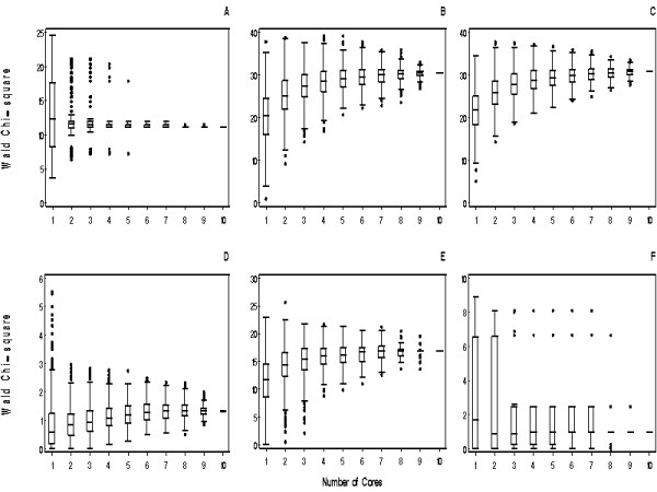
Methods: A simulated TMA with between 1 and 10 cores was designed to study tumor expression of 6 biomarkers with varied expression patterns (B7-H1, B7-H3, survivin, Ki-67, CAIX, and IMP3) using 100 patients with clear cell renal cell carcinoma (RCC). We evaluated agreement between whole tissue section and TMA immunohistochemical biomarker quantification to assess how many TMA cores are necessary to adequately represent RCC whole tissue section expression. Additionally, we evaluated associations of whole tissue section and TMA expression with RCC-specific death.
Results: The number of simulated TMA cores necessary to adequately represent whole tissue section quantification is biomarker specific. Although 2-3 cores appeared adequate for B7-H3, Ki-67, CAIX, and IMP3, even as many as 10 cores resulted in poor agreement for B7-H1 and survivin compared to RCC whole tissue sections. While whole tissue section B7-H1 was significantly associated with RCC-specific death, no significant associations were detected using as many as 10 TMA cores, suggesting that TMAs can result in false-negative findings if the TMA is not optimally designed.
Conclusions: Prior to TMA analysis, the number of TMA cores necessary to accurately represent biomarker expression on whole tissue sections should be established as there is not a one-size-fits-all TMA. We illustrate the use of a simulated TMA as a cost-effective tool for this purpose.
Figures
References
Publication types
MeSH terms
Substances
Grants and funding
LinkOut - more resources
Full Text Sources
Medical
Research Materials
