

Review
doi: 10.1016/j.chembiol.2010.04.013.
Synthetic strategies for studying embryonic development
Affiliations
- PMID: 20609409
- PMCID: PMC2902240
- DOI: 10.1016/j.chembiol.2010.04.013
Item in Clipboard
Review
Synthetic strategies for studying embryonic development
Chem Biol.
.
Abstract
Developmental biology has evolved from a descriptive science to one based on genetic principles and molecular mechanisms. Although molecular biology and genetic technologies have been the primary drivers of this transformation, synthetic strategies have been increasingly utilized to interrogate the mechanisms of embryonic patterning with spatial and temporal precision. In this review, we survey how chemical tools and engineered proteins have been used to perturb developmental processes at the DNA, RNA, protein, and cellular levels. We discuss the design principles, experimental capabilities, and limitations of each method, as well as future challenges for the chemical and developmental biology communities.
Figures
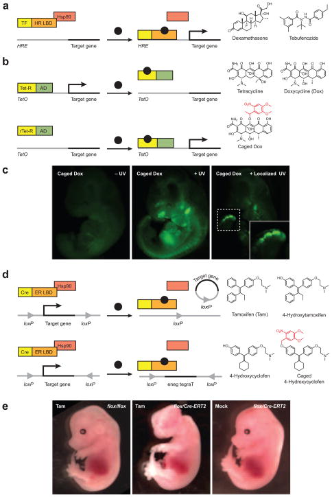
(a) Schematic representation of hormone receptor-based transactivator systems. Chemical structures for representative agonists are shown. TF, transcription factor; HR LBD, hormone receptor ligand-binding domain; HRE, hormone response element. (b) Schematic representation of tetracycline repressor-based transcription systems, including the “Tet-Off” and “Tet-On” configurations. Chemical structures of tetracycline and its synthetic derivatives are shown. Tet-R, Tet repressor; rTet-R, reverse Tet transactivator; AD, activation domain; TetO, Tet repressor operon. Although hormone receptor-and tetracycline repressor-based transactivators can each form protein dimers, monomeric forms of these proteins are depicted for schematic simplicity. (c) Control of GFP expression in mice embryos using the “Tet-On” system and caged doxycycline. Both global and localized (dashed box and inset) irradiation are shown. Adapted with permission (Cambridge et al., 2009; Copyright 2009, Nature America, Inc). (d) Schematic representation of tamoxifen-induced, Cre-dependent recombination, including excision and inversion reactions. The recombinase homodimer is depicted as a monomer for schematic simplicity, and chemical structures of tamoxifen and its derivatives are shown. ER LBD, estrogen receptor ligand-binding domain. (e) Tamoxifen-induced “self knockout” of the Otx2 gene in mouse embryos using the Cre-ERT2 system. Abnormal craniofacial and brain development is observed only in Otx2flox/Cre-ERT2 embryos treated with the ER agonist. Adapted with permission (Fossat et al., 2006; Copyright 2006, European Molecular Biology Organization).
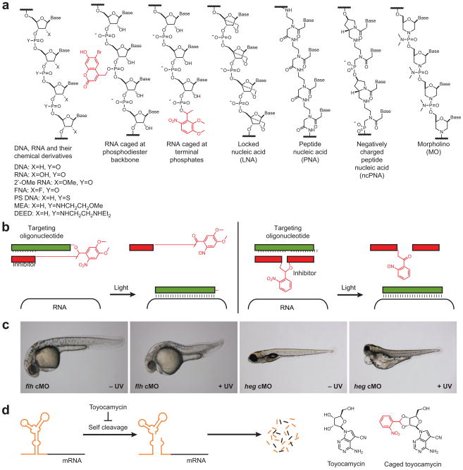
(a) Chemical structures for natural oligonucleotides, their modified analogs, and non-natural oligonucleotides. Caging groups are shown in red. (b) Schematic representation of the caged MO hairpin (left) and PhotoMorphs™ technologies (right). (c) Light-controlled silencing of flh and heg in zebrafish embryos using caged MO hairpins. Adapted with permission (Ouyang et al., 2009; Copyright 2009, American Chemical Society). (d) Schematic representation of a small molecule-dependent riboswitch (orange) that regulates transcript stability. Chemical structures of toyocamycin and its caged derivative are shown.
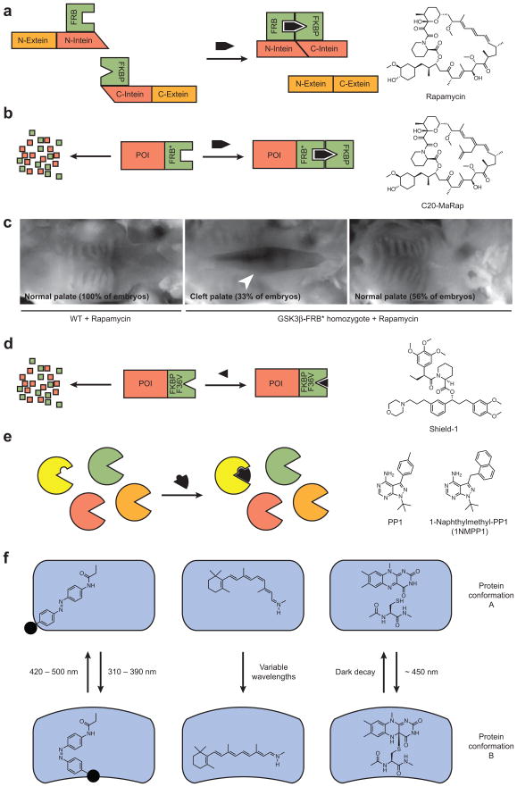
(a) Schematic representation of rapamycin-induced polypeptide splicing using fusion proteins composed of FRB, FKBP, and split inteins. (b) Schematic representation of ligand- and FKBP-dependent stabilization of FRB*-containing fusion proteins and the chemical structure of the FRB*-specific rapamycin analog C20-MaRap. POI, protein of interest. (c) Rapamycin-dependent rescue of GSK3β activity in homozygous GSK3β-FRB* knock-in mouse embryos. Rapamycin has no effect on wildtype embryos during the two-day drug regimen, but the majority of GSK3β-FRB* mice are fully rescued from cleft palate defects (white arrowhead) upon rapamycin treatment. Partial rescues were also observed (data not shown). Adapted with permission (Liu et al., 2007; Copyright 2007, Nature Publishing Group). (d) Schematic representation of ligand-dependent stabilization of FKBP mutant-containing fusion proteins and the chemical structure of Shield-1. (e) Schematic representation of the “bump-and-hole” strategy for achieving targeted protein inhibition with genome-wide specificity. Chemical structures of promiscuous (PP1) and mutant kinase-specific (naphthylmethyl-PP1) inhibitors are shown. (f) Schematic representation of light-regulated protein conformation and function using synthetic and endogenous cofactors. Azobenzene-, retinal-, and flavin mononucleotide-based approaches are shown, with the azobenzene-linked small-molecule modulator depicted as a solid black circle.
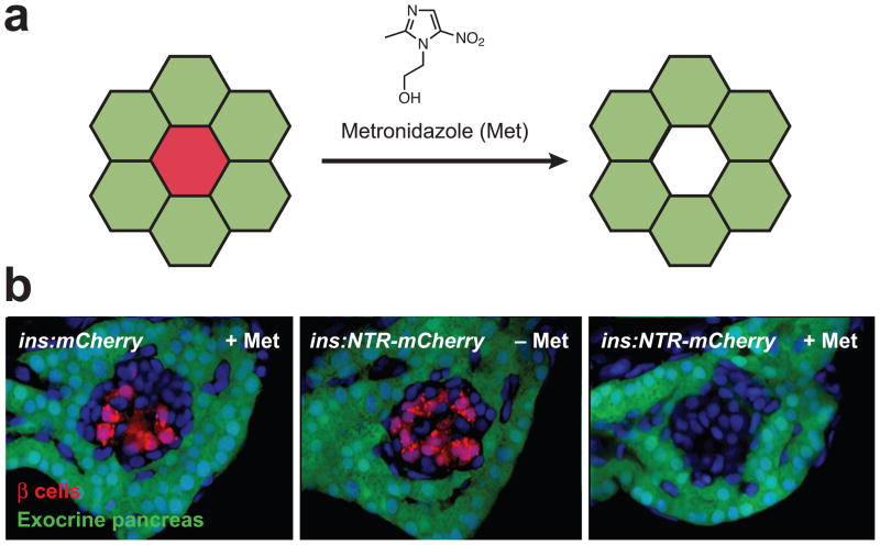
(a) Schematic representation of metronidazole-dependent apoptosis of nitroreductase-expressing cells (red) without collateral damage to adjacent cells (green). (b) Metronidazole-induced ablation of pancreatic β cells (red) in transgenic zebrafish larvae with insulin promoter-driven nitroreductase-mCherry expression (ins:NTR-mCherry). Exocrine pancreas cells (green) were not affected, and no drug-dependent cell ablation was observed in ins:mCherry larvae. Adapted with permission (Pisharath et al., 2007; Copyright 2006, Elsevier Ireland Ltd.).
References
-
- Airan RD, Thompson KR, Fenno LE, Bernstein H, Deisseroth K. Temporally precise in vivo control of intracellular signalling. Nature. 2009;458:1025–1029. - PubMed
-
- Ando H, Furuta T, Tsien RY, Okamoto H. Photo-mediated gene activation using caged RNA/DNA in zebrafish embryos. Nat Genet. 2001;28:317–325. - PubMed
Publication types
MeSH terms
Substances
Grants and funding
LinkOut - more resources
Full Text Sources
Other Literature Sources
