Review
. 2011 Feb-Mar;90(2-3):198-204.
doi: 10.1016/j.ejcb.2010.05.009. Epub 2010 Jul 6.

Role of WASP in cell polarity and podosome dynamics of myeloid cells

Affiliations
Review

Role of WASP in cell polarity and podosome dynamics of myeloid cells

James Monypenny et al. Eur J Cell Biol. 2011 Feb-Mar.

Abstract

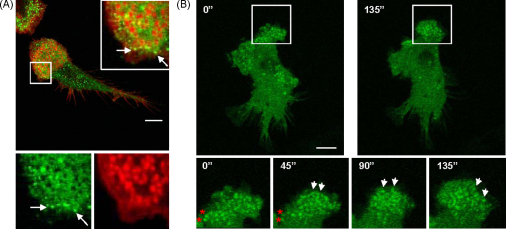
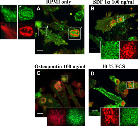
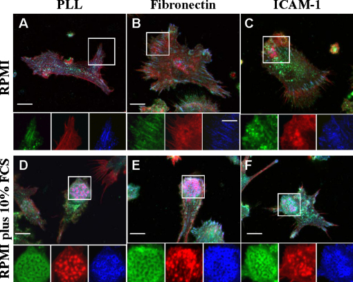
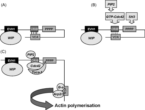
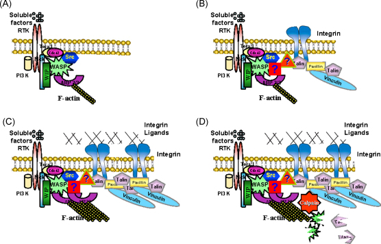
The integrin-dependent migration of myeloid cells requires tight coordination between actin-based cell membrane protrusion and integrin-mediated adhesion to form a stable leading edge. Under this mode of migration, polarised myeloid cells including dendritic cells, macrophages and osteoclasts develop podosomes that sustain the extending leading edge. Podosome integrity and dynamics vary in response to changes in the physical and biochemical properties of the cell environment. In the current article we discuss the role of various factors in initiation and stability of podosomes and the roles of the Wiskott Aldrich Syndrome Protein (WASP) in this process. We discuss recent data indicating that in a cellular context WASP is crucial not only for localised actin polymerisation at the leading edge and in podosome cores but also for coordination of integrin clustering and activation during podosome formation and disassembly.


Figures

Fig. 1
Fig. 1
Composition and dynamics of the leading edge in DCs. (A) Confocal micrograph of spleen derived mouse DCs plated on poly-l-lysine coated coverslips overnight. DCs were fixed with 3% paraformaldehyde, permeabilised with 0.05% Triton X-100 and double-stained to detect the distribution of β2 integrin (green) and actin (red). Magnifications of the boxed area show the distribution of β2 integrins and actin separately and merged. β2 Integrins form circular arrays surrounding actin puncta in podosomes and also cluster forming focal contacts at the edge of the leading edge (arrows). (B) Still images of WASP −/− spleen derived mouse DCs expressing eGFP-WASP filmed live using by confocal microscopy taking frames every 15 s. DC were plated on poly-l-lysine coated coverslips overnight and mounted onto viewing chambers. Digits show elapsed time in seconds from the beginning of the film. Magnifications of the boxed area at the bottom of the images show the distribution of eGFP-WASP at the leading edge. eGFP-WASP signal shows the dynamics of podosome assembly and disassembly. White arrows show podosomes that form or mature with respect to the previous time point. Red asterisks point at podosomes that disassemble with respect to the previous time point. As the leading edge extends, podosomes form and mature at the front (white arrows) while at the back of the podosome cluster podosomes disassemble (red asterisks). Bar 10 μm.
Fig. 2
Fig. 2
Soluble factors induce DC polarisation and podosome formation. Spleen derived mouse DCs were collected from cultures in RPMI supplemented with 10% FCS, GM-CSF and TGFβ1 and washed 3 times with RPMI to discard supplemented soluble factors. DCs were then resuspended in RPMI (A) or RPMI supplemented with 100 ng/ml SDF1α (B), 100 ng/ml osteopontin (C) or 10% Foetal Calf Serum (FCS) (D) and allowed to spread on poly-l-lysine coated coverslips for 4 h. DCs were fixed with 3% paraformaldehyde, permeabilised with 0.05% Triton X-100 and double-stained to detect the distribution of β2 integrin (green) and actin (red). Images show the merged confocal images of the distribution of both β2 integrin and actin. Magnifications of the boxed areas on the left or at the bottom of the images show the distribution of β2 integrins and actin separately. The majority of DCs plated with RPMI only developed β2 integrin containing focal contacts or rosettes of podosomes (A). SDF1α, osteopontin and FCS promoted cell polarisation (elongated morphology with a distinct leading edge sustained by a cluster of podosomes) and formation of regular clusters of podosomes with the characteristic honeycomb distribution of β2 integrins surrounding actin puncta (B–D). Bar 10 μm.
Fig. 3
Fig. 3
Integrin ligands promote accumulation of integrins and integrin-associated proteins in podosomes. Spleen derived mouse DCs were collected from cultures in RPMI supplemented with 10% FCS, GM-CSF and TGFβ1, washed 3 times with RPMI to discard supplemented soluble factors. DCs were then resuspended in RPMI (A–C) or RPMI supplemented with 10% FCS (D–F) and allowed to spread on poly-l-lysine (10 μg/ml), fibronectin (10 μg/ml) or ICAM-1 (10 μg/ml) coated coverslips for 4 h. DCs were fixed with 3% paraformaldehyde, permeabilised with 0.05% Triton X-100 and co-stained to detect the distribution of β2 integrin (green), actin (red) and vinculin (blue). Images show the merged confocal images of the distribution of β2 integrin, actin and vinculin. Magnifications of the boxed areas at the bottom of the images show the distribution of β2 integrins, actin and vinculin separately. Integrin ligands in the absence of soluble factors failed to induce cell polarisation and formation of podosome clusters behind leading edges. In the presence of soluble factors from FCS, integrin ligands induced accumulation of β2 integrins and vinculin in podosome rings. Bar 10 μm.
Fig. 4
Fig. 4
Regulation of WASP activation. EVH1: Ena/Vasp homology 1 domain; CRIB: Cdc42 and Rac interactive domain; pppp: proline rich domains; VCA: verprolin cofilin homology domains/acidic region. (A) In the inactive state, WASP adopts an auto-inhibitory conformation mediated through the interaction between the VCA and the CRIB domains. WIP is bound to the EVH1 domain when WASP is in this conformation. (B) Following cell stimulation, various factors can promote the open conformation of WASP by disturbing the interaction between the VCA and the CRIB domain. These include binding of: (i) GTP-bound Cdc42 to the CRIB domain and (ii) SH3 domain containing proteins (adaptors and kinases) to the proline-rich domain. Given that binding of PIP2 is also required for N-WASP activation and the similarities between these two proteins from the same family, it is likely that PIP2 binding is also required for WASP activation although there is no experimental evidence to support this assumption. (C) Toca-1 binds to both Cdc42 and WIP-WASP complex through the SH3 domain of Toca-1. It is not known if Toca-1 binds directly to WIP, nor experimentally determined whether WIP remains associated with WASP after WASP activation. Once WASP is activated, the VCA domain is released for Arp2/3 activation and actin monomer (G-a) binding, leading to actin polymerisation.
Fig. 5
Fig. 5
WASP and WIP are recruited simultaneously to nascent podosomes. The monocytic cell line THP-1 was transduced with eGFP-WASP and WIP-mCherry. Clones co-expressing eGFP-WASP and WIP-mCherry were obtained and plated on fibronectin coated coverslips in RPMI supplemented with FCS and 1 ng/ml TGF-β1 overnight and mounted onto viewing chambers. Under these conditions THP-1 cells assemble podosomes similarly to the treatment with TPA (Tsuboi, 2006), which makes them a very useful cellular model to study formation and dynamics of these adhesions. Cells were filmed live using by confocal microscopy taking frames every 15 s. Digits show elapsed time in seconds from the beginning of the film. Yellow colour in panels A–C indicates co-localisation of eGFP (green) in panels D–F and mCherry signals (red) in panels G–I. White arrows point at nascent podosomes in frame taken at the beginning of the film (time 0 s) that increase in size and WASP and WIP content 120 s later. Red asterisks point at newly assembled podosomes with respect to the previous time point. As the leading edge extends WASP and WIP are recruited simultaneously to the core of nascent podosomes suggesting these proteins are constituents of the podosome initiation complex. Bar 10 μm.
Fig. 6
Fig. 6
Proposed model of the role of WASP in podosome formation and dissolution. (A) Soluble factors bind to their corresponding receptors which are likely to be receptor tyrosine kinases (RTK) as the ones illustrated in the figure or G-coupled protein receptors, and lead to recruitment and association of PI3K, Cdc42 and WASP and WIP in a complex perhaps linked to other adaptor proteins such as Nck. Additionally, GEFs are recruited leading to activation of Cdc42 and likely Toca 1 is also recruited leading to WASP activation and Arp2/3-mediated actin polymerisation. (B) Simultaneously, integrins are recruited to nascent actin cores in a process where WASP and WIP work as a functional unit to bridge forming actin filaments and integrins leading to formation of the characteristic circular arrays of integrins and integrin-associated proteins surrounding the podosome cores. Other molecules that may provide the link between the WASP-WIP functional unit, F-actin and integrins remain unknown and are symbolised in the diagram with question marks. (C) If integrin ligands are present in the vicinity of the cell, recruited integrins will engage and lead to a feed back loop of activation of PI3K and Cdc42 leading to further WASP activation, actin polymerisation and recruitment of integrins resulting in increased integrity and stabilisation of podosomes. (D) Once podosomes reach a critical size and locate at the back of the cluster, calpain is activated and in parallel WASP and talin (and perhaps other still unidentified podosomal components) may become more sensitive to calpain-mediated cleavage resulting in dissolution of podosomes. Some of the drawings used in these diagrams where obtained from the publication of DeMali and Burridge (2003) with permission from the authors.

Similar articles

Cited by

References

    1. Anceriz N., Vandal K., Tessier P.A. S100A9 mediates neutrophil adhesion to fibronectin through activation of beta2 integrins. Biochem. Biophys. Res. Commun. 2007;354:84–89. - PMC - PubMed
    1. Ancliff P.J., Blundell M.P., Cory G.O., Calle Y., Worth A., Kempski H., Burns S., Jones G.E., Sinclair J., Kinnon C., Hann I.M., Gale R.E., Linch D.C., Thrasher A.J. Two novel activating mutations in the Wiskott–Aldrich syndrome protein result in congenital neutropenia. Blood. 2006;108:2182–2189. - PubMed
    1. Anton I.M., Jones G.E. WIP: a multifunctional protein involved in actin cytoskeleton regulation. Eur. J. Cell. Biol. 2006;85:295–304. - PubMed
    1. Anton I.M., Jones G.E., Wandosell F., Geha R., Ramesh N. WASP-interacting protein (WIP): working in polymerisation and much more. Trends Cell. Biol. 2007;17:555–562. - PubMed
    1. Blundell M.P., Bouma G., Calle Y., Jones G.E., Kinnon C., Thrasher A.J. Improvement of migratory defects in a murine model of Wiskott–Aldrich syndrome gene therapy. Mol. Ther. 2008;16:836–844. - PubMed

Publication types

Substances

LinkOut - more resources