

Protocols for the detection of s-glutathionylated and s-nitrosylated proteins in situ
- PMID: 20609917
- PMCID: PMC3113509
- DOI: 10.1016/S0076-6879(10)74017-9
Protocols for the detection of s-glutathionylated and s-nitrosylated proteins in situ
Abstract
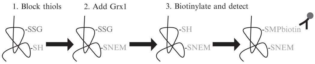
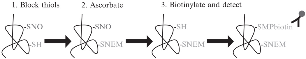
The oxidation of protein cysteine residues represents significant posttranslational modifications that impact a wide variety of signal transduction cascades and diverse biological processes. Oxidation of cysteines occurs through reactions with reactive oxygen as well as nitrogen species. These oxidative events can lead to irreversible modifications, such as the formation of sulfonic acids, or manifest as reversible modifications such as the conjugation of glutathione with the cysteine moiety, a process termed S-glutathionylation (also referred to as S-glutathiolation, or protein mixed disulfides). Similarly, S-nitrosothiols can also react with the thiol group in a process known as S-nitrosylation (or S-nitrosation). It is the latter two events that have recently come to the forefront of cellular biology through their ability to reversibly impact numerous cellular processes. Herein we describe two protocols for the detection of S-glutathionylated or S-nitrosylated proteins in situ. The protocol for the detection of S-glutathionylated proteins relies on the catalytic specificity of glutaredoxin-1 for the reduction of S-glutathionylated proteins. The protocol for the detection of S-nitrosylated proteins represents a modification of the previously described biotin switch protocol, which relies on ascorbate in the presence of chelators to decompose S-nitrosylated proteins. These techniques can be applied in situ to elucidate which compartments in tissues are affected in diseased states whose underlying pathologies are thought to represent a redox imbalance.
Copyright (c) 2010 Elsevier Inc. All rights reserved.
Figures
References
-
- Benhar M, et al. Protein denitrosylation: Enzymatic mechanisms and cellular functions. Nat. Rev. Mol. Cell Biol. 2009;10:721–732. - PubMed
-
- Chrestensen CA, et al. Acute cadmium exposure inactivates thioltransferase (Glutaredoxin), inhibits intracellular reduction of protein-glutathionyl-mixed disulfides, and initiates apoptosis. J. Biol. Chem. 2000;275:26556–26565. - PubMed
-
- Ckless K, et al. In situ detection and visualization of S-nitrosylated proteins following chemical derivatization: Identification of Ran GTPase as a target for S-nitrosylation. Nitric Oxide. 2004;11:216–227. - PubMed
MeSH terms
Substances
Grants and funding
LinkOut - more resources
Full Text Sources
Miscellaneous
