Review
. 2010 Jul-Aug;28(4):429-40.
doi: 10.1016/j.urolonc.2010.04.008.

Molecular genetics of bladder cancer: Emerging mechanisms of tumor initiation and progression

Affiliations
Review

Molecular genetics of bladder cancer: Emerging mechanisms of tumor initiation and progression

David J McConkey et al. Urol Oncol. 2010 Jul-Aug.

Abstract

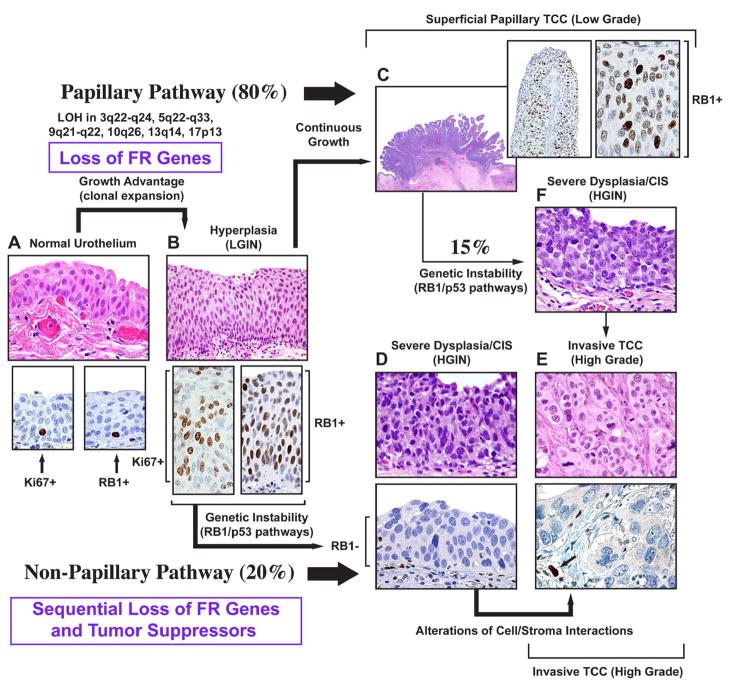
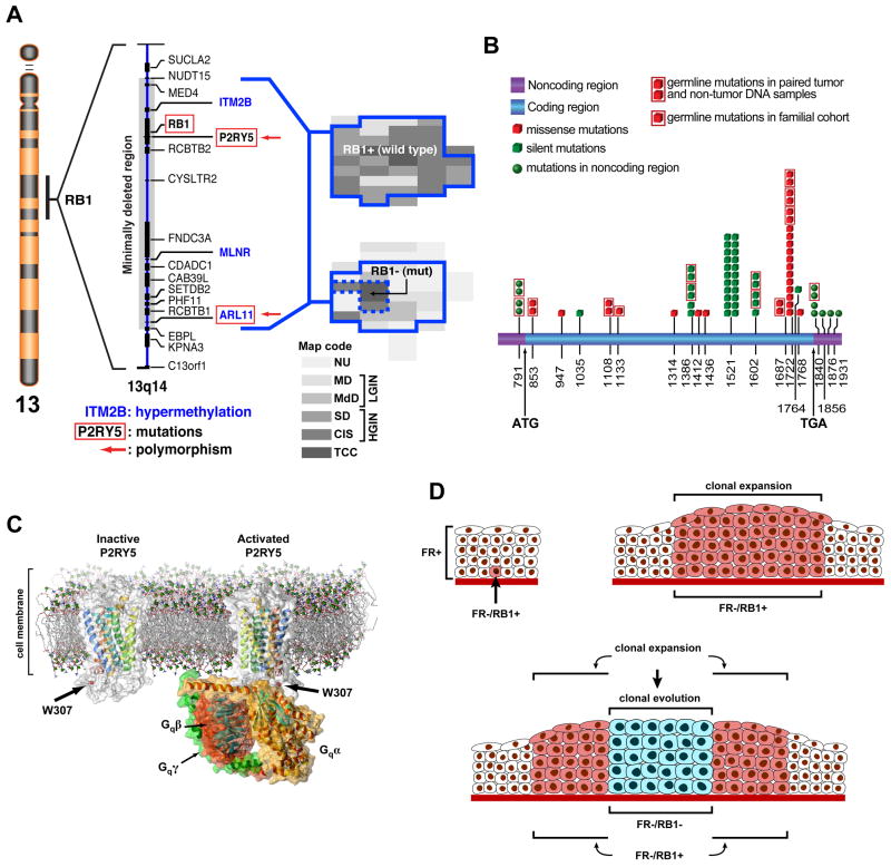
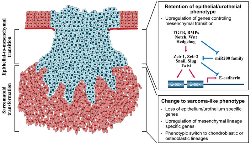
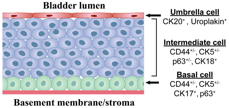
Urothelial cancer has served as one of the most important sources of information about the mutational events that underlie the development of human solid malignancies. Although "field effects" that affect the entire bladder mucosa appear to initiate disease, tumors develop along 2 distinct biological "tracks" that present vastly different challenges for clinical management. Recent whole genome methodologies have facilitated even more rapid progress in the identification of the molecular mechanisms involved in bladder cancer initiation and progression. Specifically, whole organ mapping combined with high resolution, high throughput SNP analyses have identified a novel class of candidate tumor suppressors ("forerunner genes") that localize near more familiar tumor suppressors but are disrupted at an earlier stage of cancer development. Furthermore, whole genome comparative genomic hybridization (CGH) and mRNA expression profiling have demonstrated that the 2 major subtypes of urothelial cancer (papillary/superficial and non-papillary/muscle-invasive) are truly distinct molecular entities, and in recent work our group has discovered that muscle-invasive tumors express molecular markers characteristic of a developmental process known as "epithelial-to-mesenchymal transition" (EMT). Emerging evidence indicates that urothelial cancers contain subpopulations of tumor-initiating cells ("cancer stem cells") but the phenotypes of these cells in different tumors are heterogeneous, raising questions about whether or not the 2 major subtypes of cancer share a common precursor. This review will provide an overview of these new insights and discuss priorities for future investigation.


Figures

Figure 1
Figure 1. Dual-track concept of bladder carcinogenesis
The expansion of a preneoplastic clone which shows minimal phenotypic deviation from the normal urothelium, is the incipient event in bladder carcinogenesis referred to as LGIN. In this phase, the loss of FR genes function provides growth advantage associated with the expansion of proliferating compartment. The proliferating cells expressing normal RB protein are seen in the entire thickness of LGIN. In contrast, normal urothelium contains only scattered proliferating cells expressing RB protein located in its basal layer. The continuous growth of LGIN leads to the development of low grade superficial papillary TCC. In the non-papillary pathway, clonal evolution results in the establishment of a successor clone with microscopic features of HGIN which often shows a loss of major tumor suppressors such as RB1 and has a high propensity for progression to an invasive high grade non papillary TCC. a, Normal urothelium (upper panel). Expression of Ki67 in proliferating basal cells of normal urothelium (lower panel, left). Expression of RB protein in peribasal cells of normal urothelium (lower panel, right). b, Urothelial hyperplasia with mild atypia referred to as LGIN (upper panel). Expression of Ki67 in the entire thickness of LGIN (lower panel, left); expression of RB protein in the entire thickness of LGIN (lower panel, right). c, Low-grade superficial TCC retaining normal expression of the RB protein: insets to c show low and high power photomicrographs illustrating the expression of normal RB protein in low grade papillary TCC. d, Severe intraurothelial dysplasia/carcinoma in situ (HGIN) (upper panel). Loss of RB protein expression in HGIN (lower panel). e, High-grade invasive nonpapillary carcinoma (upper panel). Loss of RB protein expression in high grade invasive nonpapillary TCC. Arrow shows expression of RB protein in endothelial cells adjacent to tumor which serve as an internal positive control (lower panel). f, Severe intraurothelial dysplasia/carcinoma in situ developing in bladder mucosa adjacent to a low-grade papillary tumor. It is responsible for switching the pathway and progression of some low-grade papillary tumors to high-grade invasive cancers. (Modified and reprinted with permission from T. Majewski et al. Lab Invest 88:694–721, 2008).
Figure 2
Figure 2. The concept of FR genes
a, Chromosome 13, shown on the left, with the expanded 13q14 region flanking RB1 depicts a deleted segment associated with early expansion of in situ bladder neoplasia and containing candidate FR genes. The candidate FR genes inactivated by nucleotide substitutions (mutations or polymorphism) are depicted by red boxes. The candidate FR genes inactivated by hypermethylation are printed in blue. Histologic maps of human cystectomies with invasive bladder cancer are shown on the right. The continuous blue line depicts plaque like areas of clonal intraurothelial expansion associated with the loss of hetrozygosity within the minimal deleted region around RB1. The upper map shows a plaque-like clonal expansion involving almost the entire bladder mucosa with no inactiviation of the remaining RB1 allele. The lower map shows the inactivation of the remaining RB1 allele by a mutation restricted to an area of invasive cancer and adjacent HGIN depicted by a dashed blue line. This mutation involved codon 556 of exon 17 consisting of CGA→TGA and resulting in the change of Arg to a stop codon. b Summary of sequence analysis of P2RY5. The positions of nucleotide substitutions are shown on the full length mRNA. c, A model of inactive P2RY5 containing 7 transmembrane (H1–H7) and one cytoplasmic (H8) helix structures showing the position of polymorphism in codon 307 located within the cystoplasmic domain of the protein (left diagram) that may affect its interaction with the Gαβγ trimeric protein complex (right diagram). d, Expansion of intraurothelial neoplasia and its clonal evolution into carcinoma in situ. Inactivation of the FR genes (FR−) in a parabasal urothelial tumor initiating cell with the retention of normal RB1 expression (RB1+) (upper left panel). Clonal expansion of the FR−/RB1+ cell shown in a exhibiting minimal deviation from a normal phenotype. Typically in this phase only an increased number of urothelial cell layers can be identified in the urothelium. (upper right panel) Clonal evolution into carcinoma in situ associated with loss of RB1 function (RB1−). (bottom panel) (a, b, and c modified and reprinted with permission from S. Lee et al PNAS 104(34):13732–13737, 2007).
Figure 3
Figure 3. Molecular regulation of epithelial-to-mesenchymal transition and sarcomatoid transformation
Some conventional bladder carcinomas develop a gene expression profile characteristic of EMT but microscopically they retain a full epithelial/urothelial phenotype. Further progression to sarcomatoid transformation is associated with the partial or complete loss of epithelial phenotype and the development of mesenchymal sarcoma-type features. The hallmark feature of EMT is a loss of the homotypic adhesion molecule, E-cadherin, which is the canonical marker of an “epithelial” phenotype. E-cadherin transcription is controlled by two E-box elements located within its promoter. EMT can be induced by a variety of developmental signals, but transforming growth factor-beta (TGFb) is the best-studied stimulus. TGFβ upregulates several E-cadherin repressors (Zeb-1, Zeb-2, Snail, Twist) that inhibit E-cadherin expression by recruiting histone deacetylases (HDACs) to the E-box elements in its promoter. In addition, TGFβ downregulates expression of the miR200 family of micro-RNAs. The miR200 family interacts directly with the mRNAs encoding Zeb-1 and Zeb-2, thereby blocking their translation and promoting their degradation. In urothelial cancers expression of ΔN-p63 correlates directly with E-cadherin expression and inversely with expression of Zeb-1 and Zeb-2. Superficial urothelial tumors almost invariably display an “epithelial” phenotype, whereas muscle-invasive tumors are heterogeneous and approximately evenly divided between “epithelial” and “mesenchymal” phenotypes. Change to sarcoma-type phenotype is relatively rare and occurs in less than 10% of high grade invasive bladder cancers. Such tumors are characterized by complex chromosomal abnormalities, pronounced aneuploidy and a high degree of clinical aggressiveness.
Figure 4
Figure 4. Differentiation-based lineage hierarchy in the normal urothelium
The normal urothelium is comprised of 3 distinct layers. The surface epithelium consists of a single layer of terminally differentiated urothelial cells (“umbrella cells”) that express uroplakins and cytokeratin-20 (CK20) and have a low proliferative potential. It is conceivable that papillomas arise from transformation of umbrella cells, but this would require that they “de-differentiate” to reacquire self-renewal potential. Below the umbrella cells is a multi-cellular layer of intermediate cells that express CK18 and variable levels of p63, CK5, and CD44. These are most likely “transit-amplifying cells” that possess higher proliferative potential but have also begun to differentiate towards the umbrella cell phenotype. Finally, adjacent to the basement membrane is a layer of basal cells that express high levels of CK5, CD44, p63 and the 67 kD laminin receptor. This layer contains urothelial stem cells, which are tightly regulated by stromal elements (cells and matrix proteins) contained within the “niche”. Recent studies indicate that urothelial cancer stem cells share several molecular markers with the normal basal cell, including CD44, CK5, and the 67 kD laminin receptor and that these markers are upregulated at the tumor-stromal interface in xenografts.

References

    1. Dinney CP, McConkey DJ, Millikan RE, et al. Focus on bladder cancer. Cancer Cell. 2004;6:111–6. - PubMed
    1. Olumi AF, Tsai YC, Nichols PW, et al. Allelic loss of chromosome 17p distinguishes high grade from low grade transitional cell carcinomas of the bladder. Cancer Res. 1990;50:7081–3. - PubMed
    1. Tsai YC, Nichols PW, Hiti AL, Williams Z, Skinner DG, Jones PA. Allelic losses of chromosomes 9, 11, and 17 in human bladder cancer. Cancer Res. 1990;50:44–7. - PubMed
    1. Ruppert JM, Tokino K, Sidransky D. Evidence for two bladder cancer suppressor loci on human chromosome 9. Cancer Res. 1993;53:5093–5. - PubMed
    1. Miyao N, Tsai YC, Lerner SP, et al. Role of chromosome 9 in human bladder cancer. Cancer Res. 1993;53:4066–70. - PubMed