. 2010 Dec 1;88(3):471-81.
doi: 10.1093/cvr/cvq218. Epub 2010 Jul 7.

Thrombospondin-1 supports blood pressure by limiting eNOS activation and endothelial-dependent vasorelaxation

Affiliations

Thrombospondin-1 supports blood pressure by limiting eNOS activation and endothelial-dependent vasorelaxation

Eileen M Bauer et al. Cardiovasc Res. .

Abstract

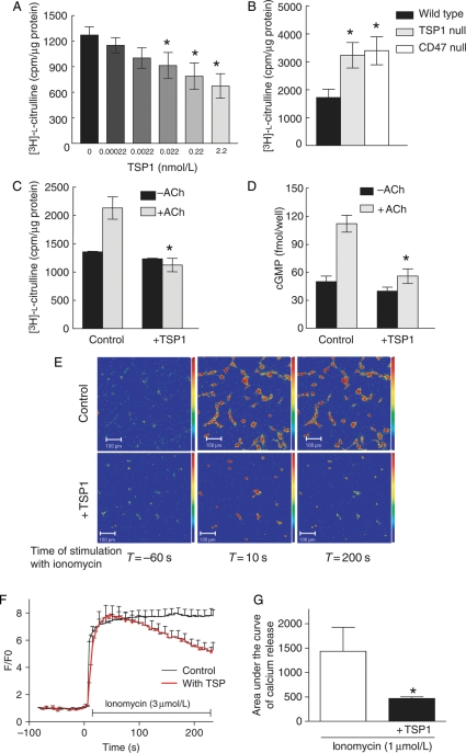
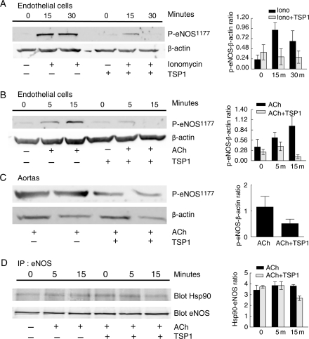
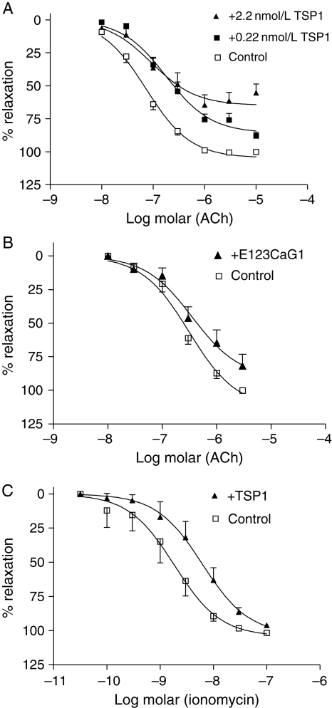
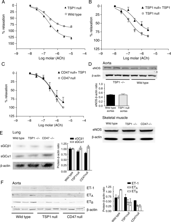
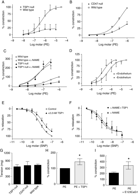
Aims: Thrombospondin-1 (TSP1), via its necessary receptor CD47, inhibits nitric oxide (NO)-stimulated soluble guanylate cyclase activation in vascular smooth muscle cells, and TSP1-null mice have increased shear-dependent blood flow compared with wild-type mice. Yet, the endothelial basement membrane should in theory function as a barrier to diffusion of soluble TSP1 into the arterial smooth muscle cell layer. These findings suggested that endothelial-dependent differences in blood flow in TSP1-null mice may be the result of direct modulation of endothelial NO synthase (eNOS) activation by circulating TSP1. Here we tested the hypothesis that TSP1 inhibits eNOS activation and endothelial-dependent arterial relaxation.

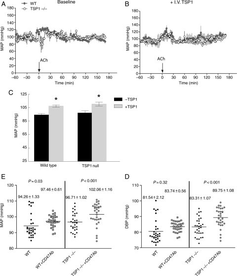
Methods and results: Acetylcholine (ACh)-stimulated activation of eNOS and agonist-driven calcium transients in endothelial cells were inhibited by TSP1. TSP1 also inhibited eNOS phosphorylation at serine(1177). TSP1 treatment of the endothelium of wild-type and TSP1-null but not CD47-null arteries inhibited ACh-stimulated relaxation. TSP1-null vessels demonstrated greater endothelial-dependent vasorelaxation compared with the wild type. Conversely, TSP1-null arteries demonstrated less vasoconstriction to phenylephrine compared with the wild type, which was corrected upon inhibition of eNOS. In TSP1-null mice, intravenous TSP1 blocked ACh-stimulated decreases in blood pressure, and both intravenous TSP1 and a CD47 agonist antibody acutely elevated blood pressure in mice.

Conclusion: TSP1, via CD47, inhibits eNOS activation and endothelial-dependent arterial relaxation and limits ACh-driven decreases in blood pressure. Conversely, intravenous TSP1 and a CD47 antibody increase blood pressure. These findings suggest that circulating TSP1, by limiting endogenous NO production, functions as a pressor agent supporting blood pressure.


Figures

Figure 1
Figure 1
TSP1 inhibits basal- and agonist-stimulated eNOS activity and modulates calcium transients. BAEC or wild-type, TSP1-null, and CD47-null endothelial cells (5 × 10⁵ cells/well) were serum starved for 24 h and incubated in minimal medium with the indicated amounts of TSP1 (A), without any treatments (B) or with TSP1 (2.2 nmol/L) followed by ACh stimulation (10 µmol/L) (C) in l-arginine free minimal medium prior to adding [3H]-l-arginine. [3H]-l-citrulline synthesis was determined as described and is presented normalized to total protein. Treatment wells were done in triplicate and all experiments were repeated three times. *P< 0.05 compared with untreated (A), wild-type (B), or ACh alone (C). HUVEC (5 × 10⁵ cells/well) were serum starved for 24 h and incubated in serum/additive free medium + 0.1% BSA and stimulated with ACh (10 µmol/L) ± TSP1 (2.2 nmol/L). Intracellular cGMP levels were determined using an enzyme immunoassay (D). HUVEC were serum starved in basal medium overnight, loaded with fluo-4-AM for 20 min, and then pre-treated with/without TSP1 (2.2 nmol/L) followed by ionomycin (3 µmol/L). Images were acquired every 3 s (×20, 259 × 259). Data analysis was performed using Pascal 3.2 software (LSM5 Pascal Zeiss) (E). Colour images are representative of quantification in (E) (fluorescence intensity bar on right). Analysed relative changes in fluorescence intensity over time (F/F0) as the result of three independent experiments (untreated cells n = 113, TSP1-treated cells n = 64, +SD) (F). Measurement of the [Ca2+]i flux in situ in fresh aortic segments from wild-type mice was performed using fluo-4-AM following treatment with/without TSP1 (2.2 nmol/L 15 min) followed by ionomycin (3 µmol/L) (G). Results expressed as the change in area under the curve. Results are the mean ± SD of six vessels treated with ionomycin and four vessels treated with ionomycin + TSP1. *P< 0.05 compared with ionomycin alone.
Figure 2
Figure 2
TSP1 inhibits agonist-stimulated phosphorylation of eNOS. HUVEC were serum starved and pre-treated with TSP1 (2.2 nmol/L 15 min) followed by addition of ionomycin (3 µmol/L) (A) or ACh (10 µmol/L) for the times indicated (B). Lysates were prepared and western blot analysis of eNOS phosphorylation at serine1177 determined. A representative blot of three experiments for each treatment is shown. Arterial segments from wild-type mice were pre-treated with TSP1 (2.2 nmol/L 15 min) followed by ACh (10 µmol/L) ± and lysates blotted against eNOS phosphorylation at serine1177 (C). A representative blot of three separate experiments is presented. HUVEC were serum starved and pre-treated with TSP1 (2.2 nmol/L 15 min) followed by ACh (10 µmol/L) for the indicated times. eNOS was immunoprecipitated and then western blotted against Hsp90 (D). A representative blot of three separate experiments is presented. For western blots, expression was quantified by densitometry calculated by measuring intensity of bands using Image J and normalized to either β-actin or eNOS expression.
Figure 3
Figure 3
TSP1 limits agonist-stimulated vasorelaxation. The luminal endothelium of wild-type vessels was pre-treated with exogenous TSP1 [0.22 and 2.2 nmol/L, (A)] or a recombinant domain of TSP1 [E123CaG1, 2.2 nmol/L, (B)] for 15 min and vasorelaxation to ACh determined. Results are the mean ± SD of 16 control vessels, 10 TSP1 (2.2 nmol/L)-treated vessels, six TSP1 (0.22 nmol/L)-treated vessels, and five E123CaG1 domain (2.2 nmol/L)-treated vessels. P< 0.0001 for both doses of TSP1 and P< 0.0063 for recombinant domain compared with untreated. Vasorelaxation of pre-contracted aortic segments from wild-type mice was determined in the presence of ionomycin (3 µmol/L) ± TSP1 (2.2 nmol/L) (C). Results are the mean ± SD of five vessels per treatment group, P< 0.0008 compared with untreated. Maximal relaxation in control vessels was set to 100% relaxation and treated vessels normalized to untreated. All fitted curves were analysed by two-way ANOVA followed by the Bonferroni post-test.
Figure 4
Figure 4
CD47 is necessary for TSP1 inhibition of eNOS-dependent arterial relaxation. Vasorelaxation dose-response curves of wild-type and TSP1-null vessels were determined to ACh (A). Data represent the mean ± SD of six TSP1-null and four control vessels. P< 0.001 TSP1-null compared with wild-type on two-way ANOVA and Bonferroni post-test. Vasorelaxation to ACh was determined in TSP1- (B) and CD47-null (C) aortic segments before and after the treatment of the luminal endothelium with TSP1 (2.2 nmol/L). Data represent the mean ± SD of four TSP1-null vessels and five CD47-null vessels. Two-way ANOVA analysis and Bonferroni post-test of the significance of TSP1 treatment of TSP1-null arteries on ACh-stimulated vasorelaxation showed P< 0.0028 compared with untreated TSP1-null arteries. Lysates from thoracic aortic segments (n = 3 vessels per strain) and vastus medialis muscle biopsies from age- and sex-matched wild-type and null mice were probed by western analysis for total eNOS (D). For each tissue type, representative blots of three experiments are presented. Lung tissue from age- and sex-matched wild-type, TSP1- and CD47-null mice was processed for western analysis of sGC subunits α1 and β1 (E). A representative blot of three experiments is presented. Lysates from thoracic aortic segments from age-matched male wild-type, TSP1- and CD47-null mice (n = 3 animals per strain) were probed via western analysis for ETA, ETB, and ET-1 (F). For western blots, expression was quantified by densitometry calculated by measuring intensity of bands using Image J and normalized to β-actin expression.
Figure 5
Figure 5
TSP1 potentiates PE-stimulated vasoconstriction. Vasoconstriction to PE was determined in wild-type and TSP1-null arteries (A) or wild-type and CD47-null arteries (B). Data represent the mean ± SD of eight vessels from each strain (A) and 12 vessels from each strain (B). Two-way ANOVA analysis and Bonferroni post-test of significance of PE-stimulated vasoconstriction in wild-type vs. TSP1- and CD47-null arteries showed P< 0.0001. Vasoconstriction to PE±l-NAME (100 µmol/L) was determined in wild-type and TSP1-null arteries (C). Data represent the mean ± SD of five vessels of each strain. Two-way ANOVA analysis and Bonferroni post-test of significance of PE-stimulated vasoconstriction in wild-type vs. TSP1-null vessels showed P< 0.0001 compared with no difference in significance in vasoconstriction in wild-type and null vessels treated with PE + l-NAME. Changes in arterial tone to PE were determined in wild-type vessels with and without endothelium (D), in wild-type vessels with intact endothelium to SNP ± TSP1 (2.2 nmol/L) (E) and in l-NAME (100 µmol/L) treated wild-type vessels to SNP ± TSP1 (2.2 nmol/L) (F). Data represent the mean ± SD of 26 vessels with intact endothelium, 11 denuded vessels, four vessels treated with SNP, and four vessels treated with SNP + TSP1. Two-way ANOVA analysis and Bonferroni post-test of significance between endothelial intact vs. denuded vessels showed P< 0.0001 and between vessels treated with SNP vs. SNP + TSP1 showed P< 0.0003. Vasoconstriction in wild-type, TSP1-null, and CD47-null vessels was determined to KCl [100 mmol/L, (G)]. Data represent the mean ± SD of four vessels in each treatment group. P =0.510 and 0.916 wild type compared with TSP1 and CD47 null, respectively. Vasoconstriction of wild-type arteries was determined in response to an EC80 dose of PE (1 µmol/L) ± TSP1 (2.2 nmol/L) (H) or a recombinant fragment of the signature domain of TSP1 (E123CaG1, 2.2 nmol/L) (I). Data represent the mean ± SD of four vessels in each treatment group. P< 0.05 compared with PE alone (H and I).
Figure 6
Figure 6
Circulating TSP1 limits endothelial-dependent changes in blood pressure and is a hypertensive on acute administration. Age-matched male wild-type and TSP1-null mice bearing telemetric pressure transducers were treated with i.v. ACh (0.08 μg/gram weight) and MAP determined (A). TSP1-null mice were pre-treated with intravenous TSP1 (14.7 pmol/gram body weight) and followed 3 h later by i.v. ACh and MAP determined (B). Age-matched male wild-type and TSP1-null mice were treated with intravenous TSP1 (22 pmol/gram body weight) and MAP determined via telemetry transducer (C). Results represent the mean ± SD over a 2 h time interval post-administration of four animals of each strain. P< 0.05 compared with untreated. Age-matched wild-type and TSP1-null male mice received a CD47 antibody (clone 301, 4 μg/gram weight) or vehicle (normal saline) and the change in MAP and diastolic blood pressure determined via telemetric transducer. Results represent the mean ± SD over a 2 h time interval post-administration of eight animals of each strain.

Comment in

References

    1. Dudzinski DM, Michel T. Life history of eNOS: partners and pathways. Cardiovasc Res. 2007;75:247–260. doi:10.1016/j.cardiores.2007.03.023. - DOI - PMC - PubMed
    1. Dudzinski DM, Igarashi J, Greif D, Michel T. The regulation and pharmacology of endothelial nitric oxide synthase. Annu Rev Pharmacol Toxicol. 2006;46:235–276. doi:10.1146/annurev.pharmtox.44.101802.121844. - DOI - PubMed
    1. Landmesser U, Hornig B, Drexler H. Endothelial function: a critical determinant in atherosclerosis? Circulation. 2004;109:II27–II33. - PubMed
    1. Sauzeau V, Le Jeune H, Cario-Toumaniantz C, Smolenski A, Lohmann SM, Bertoglio J, et al. Cyclic GMP-dependent protein kinase signaling pathway inhibits RhoA-induced Ca2+ sensitization of contraction in vascular smooth muscle. J Biol Chem. 2000;275:21722–21729. doi:10.1074/jbc.M000753200. - DOI - PubMed
    1. Isenberg JS, Ridnour LA, Perruccio EM, Espey MG, Wink DA, Roberts DD. Thrombospondin-1 inhibits endothelial cell responses to nitric oxide in a cGMP-dependent manner. Proc Natl Acad Sci USA. 2005;102:13141–13146. doi:10.1073/pnas.0502977102. - DOI - PMC - PubMed

Publication types

MeSH terms