. 2010 Sep 1;21(17):3041-53.
doi: 10.1091/mbc.E10-04-0313. Epub 2010 Jul 7.

FIP1/RCP binding to Golgin-97 regulates retrograde transport from recycling endosomes to the trans-Golgi network

Affiliations

FIP1/RCP binding to Golgin-97 regulates retrograde transport from recycling endosomes to the trans-Golgi network

Jian Jing et al. Mol Biol Cell. .

Abstract

Many proteins are retrieved to the trans-Golgi Network (TGN) from the endosomal system through several retrograde transport pathways to maintain the composition and function of the TGN. However, the molecular mechanisms involved in these distinct retrograde pathways remain to be fully understood. Here we have used fluorescence and electron microscopy as well as various functional transport assays to show that Rab11a/b and its binding protein FIP1/RCP are both required for the retrograde delivery of TGN38 and Shiga toxin from early/recycling endosomes to the TGN, but not for the retrieval of mannose-6-phosphate receptor from late endosomes. Furthermore, by proteomic analysis we identified Golgin-97 as a FIP1/RCP-binding protein. The FIP1/RCP-binding domain maps to the C-terminus of Golgin-97, adjacent to its GRIP domain. Binding of FIP1/RCP to Golgin-97 does not affect Golgin-97 recruitment to the TGN, but appears to regulate the targeting of retrograde transport vesicles to the TGN. Thus, we propose that FIP1/RCP binding to Golgin-97 is required for tethering and fusion of recycling endosome-derived retrograde transport vesicles to the TGN.


Figures
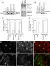
Figure 1.
Figure 1.
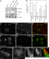
Rab11 is required for TGN38 retrograde transport to TGN. (A, left) RT-PCR analysis to determine the efficiencies of Rab11a and Rab11b knockdown. (A, right) Lysates from mock- or Rab11a/b siRNA-treated cells were immunoblotted with pan-Rab11, anti-Syntaxin 6, anti-FIP1/RCP, and anti-FIP5/Rip11 antibodies. (B) Mock- or Rab11a/b siRNA-treated cells were fractionated into cytosol and membrane fractions and immunoblotted for Rab11-binding protein FIP1/RCP and transferring receptor. Please note that in these immunoblots we show only the predominant ∼70-kDa splice isoform of FIP1/RCP. (C and D) Mock-, Rab11a, Rab11b, or Rab11a/b siRNA-treated cells HeLa-Tac-TGN38 cells were analyzed. Cells were scored for TGN38 localization and divided in dispersed or TGN-localized categories. n is the number of cells analyzed. (E–J) Mock- (D–G) or Rab11a/b siRNA-treated (H–J) HeLa-Tac-TGN38 cells were fixed and costained with anti-FIP1/RCP (red channel, F and I) and anti-Tac (green channel, E and H) antibodies.
Figure 2.
Figure 2.
FIP1/RCP knockdown affects the retrograde transport of TGN38. (A) Stable HeLa-Tac-TGN38 cells were either mock-transfected or transfected with FIP1/RCP, FIP5/Rip11, or FIP3 siRNAs. Cells were then harvested and the total levels of FIP1/RCP, FIP5/Rip11, FIP3, and Syntaxin 6 were determined by immunoblotting. Please note that although siRNAs caused the knockdown of both splice isoforms, in these immunoblots we only show the predominant ∼70-kDa splice isoforms of FIP1/RCP and FIP5/Rip11. (B) Tac-TGN38 localization was analyzed in mock-, FIP1/RCP, FIP5/Rip11, or FIP3 siRNA-treated cells. Cells were scored for TGN38 localization and divided as dispersed or TGN-localized categories. n is the number of cells analyzed. (C–J) Mock- (C–E and I) or FIP1/RCP siRNA-treated (F–H and J) HeLa-Tac-TGN38 cells were fixed and stained with anti-FIP1/RCP (D and G) and anti-Tac (C, F, I, and J) antibodies. I and J are higher-magnification images of the boxed regions from C and F. (K–M) FIP1/RCP siRNA-treated HeLa-Tac-TGN38 cells were fixed and stained with anti-Syntaxin 6 (L and M) and anti-Tac (K and M) antibodies.
Figure 3.
Figure 3.
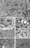
FIP1/RCP is required for the transport of TGN38 from endosomes to TGN. (A–E) HeLa cells treated with either mock siRNA or FIP1/RCP siRNA were transfected with HRP-TGN38. (A) FIP1/RCP siRNA-treated cells with panel showing mock-treated cells; scale bars, 1 μm. (B and D) FIP1/RCP siRNA-treated cells in the perinuclear region and periphery, respectively; scale bars, (B) 500 nm, (D) 1 μm. (C and E) Mock siRNA-treated cells in the perinuclear region and periphery, respectively; scale bars, (C) 500 nm, (E) 1 μm. (F and) G) HeLa cells transfected with TGN38-HRP and GFP-RBD; scale bars, (F) 500 nm, (G) 1 μm.
Figure 4.
Figure 4.
Retrograde transport of STxB is impaired in FIP1/RCP-depleted cells. (A and B) HeLa cells were incubated with 6His-STxB at 18°C for 60 min to accumulate in EE. Cells were then washed, fixed, and stained with anti-FIP1/RCP (B) and anti-6His (A) antibodies. (C–F and H–I) Mock- (C, D, and H) or FIP1/RCP (E, F, and I) siRNA-treated HeLa cells were incubated with 6His-STxB at 18°C for 60 min. Cells were then washed, and retrograde transport of 6His-STxB from EE to TGN was initiated by incubating cells at 37°C for 90 min. Cells were then fixed and stained with anti-FIP1/RCP (D and F) and anti-6His (C, E, H, and I) antibodies. H and I are higher-magnification images from the boxed regions in C and E. (G) Quantitation of 6His-STxB localization (TGN or dispersed) in mock- or FIP1/RCP siRNA-treated cells, after loading cells with 6His-STxB at 18°C, followed by 90-min incubation at 37°C as in C–I. n is the number of cells analyzed.
Figure 5.
Figure 5.
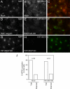
FIP1/RCP is not required for CI-MPR6 retrograde transport. (A–I) Mock- (A–C), Rab11a/b (D–F), or FIP1/RCP (G–I) siRNA-treated HeLa cells, stably expressing CD8-CI-MPR6, were fixed and stained with anti-FIP1/RCP (B, E, and H, red channel) and anti-CD8 (A, D, and G, green channel) antibodies. (J) Quantitation of CD8-CI-MPR6 localization (TGN or dispersed) in mock- or FIP1/RCP siRNA-treated HeLa cells. n is the number of cells analyzed.
Figure 6.
Figure 6.
Golgin-97 is a FIP1/RCP-binding protein. (A and B) FIP1/RCP was immunoprecipitated from HeLa cell lysate with anti-FIP1/RCP antibodies. The candidate bands in the anti-FIP1/RCP but not the nonspecific IgG lane were cut from the gel and analyzed by MS/MS-LCQ. The immunoprecipitation and subsequent proteomic analysis was done three times. Only proteins identified all three times are listed in the figure. (C) FIP1/RCP or FIP5/Rip11 were immunoprecipitated from HeLa cell lysate with anti-FIP1/RCP or anti-FIP5/Rip11 antibodies and analyzed for the presence of Golgin-97 by Western blotting with anti-Golgin-97 antibody. (D) FIP1/RCP was immunoprecipitated from HeLa cells transfected with GFP-Golgin-97. Precipitate was then analyzed for the presence of FIP1/RCP, Golgin-97, and GFP-Golgin-97.
Figure 7.
Figure 7.
Mapping the binding between Golgin-97 and FIP1/RCP. (A) The interactions between different truncation mutants of Golgin-97, FIP1/RCP, and FIP5/Rip11 were analyzed using the yeast two-hybrid assay. Growth of three separate yeast clones in −Leu/−Trp and −Leu/−Trp/−His/−Ade plates were analyzed for each binding combination. (B and C) Truncation analyses of binding between Golgin-97 and FIP1/RCP were carried out using the yeast two-hybrid assay. Three different yeast clones were analyzed for each binding combination and the results summarized as binding (+) or not (−). (D and E) To confirm the yeast two-hybrid assays, 6His-Golgin-97(aa603-767) or GST-Golgin-97(aa603-767) was expressed using the baculovirus system and purified using Ni2+ chromatography (see Coomassie gel in the bottom panel). (E, left) Purified recombinant 6His-Golgin-97(aa603-767) was incubated with glutathione beads coated with either GST alone or GST-FIP1/RCP. Beads were then washed and presence of 6His-Golgin-97(aa603-767) tested by immunoblotting with anti-His6. (E, right) Recombinant 6His-FIP1/RCP was incubated with glutathione beads coated with either GST alone or GST-Golgin-97(aa603-767). Beads were then washed and presence of 6His-FIP1/RCP tested by immunoblotting with anti-FIP1/RCP antibody.
Figure 8.
Figure 8.
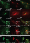
GFP-Golgin-97(aa603-767) localizes to TGN and is sufficient to mediate tac-TGN38 retrograde transport. (A–C) HeLa-Tac-TGN38 cells were transfected with GFP-Golgin-97(aa603-767). Cells were then fixed and stained with anti-Tac antibodies (B). Yellow in C represents the degree of overlap between GFP-Golgin-97(aa603-767, green) and Tac-TGN38 (red). (D–G) Mock- (D and E) or Golgin-97 siRNA-treated (F and G) cells were fixed and stained with anti-Syntaxin 6 (D and F) and anti-Golgin-97 (E and G) antibodies. Inset in E is an anti-Golgin-97 immunoblot of lysates from mock- or Golgin-97 siRNA-treated cells showing the extent of Golgin-97 knockdown. (H–J) HeLa cells stably expressing Tac-TGN38 were treated with Golgin-97 siRNA. Cells were then fixed and stained with anti-Syntaxin 6 and anti-tac antibodies. Number in H is the percentage of cells exhibiting shown phenotype. n is the number of cells analyzed. Yellow in J represents the degree of overlap between Syntaxin 6 and tac-TGN38. Arrow in high-magnification insets point to cytosolic organelles containing Syntaxin 6 and Tac-TGN38. (K–M) HeLa cells stably expressing tac-TGN38 were treated with Golgin-97 siRNA and GFP-Golgin-97(aa603-767). Cells were then fixed and stained with anti-Tac antibodies. Yellow in M represents the degree of overlap between GFP-Golgin-97(aa603-767) and Tac-TGN38.
Figure 9.
Figure 9.
TGN38 partially colocalizes with TfR, EpsinR, and retromer complex in FIP1/RCP knockdown cells. (A–C and J–L) Mock- (A–C) or FIP1/RCP siRNA-treated (J–L) HeLa-Tac-TGN38 cells were fixed and stained with anti-SNX1 (green) and anti-Tac (red) antibodies. Arrows in J–L point to organelles where TGN38 and SNX1 overlap. (D–F and M–P) Mock- (D–F) or FIP1/RCP siRNA-treated (M–P) HeLa-Tac-TGN38 cells were fixed and stained with anti-EpsinR (red) and anti-Tac (green) antibodies. Arrows in M–P point to organelles where TGN38 and EpsinR overlap. (G–I) FIP1/RCP siRNA-treated HeLa-Tac-TGN38 cells were incubated with Tf-594 (5 μg/ml) for 30 min at 37°C. Cells were then washed, fixed, and stained with anti-Tac (A and C, green) antibodies. Arrows point to organelles where TGN38 and Tf overlap.
Figure 10.
Figure 10.
Working model for FIP1/RCP function in retrograde transport. (A) FIP1/RCP is required for TGN38 and Shiga toxin retrograde transport via recycling endosomes. (B) Schematic representation of FIP1/RCP and Golgin-97 roles in tethering retrograde endocytic carriers to TGN.

Similar articles

Cited by

References

    1. Arighi C. N., Hartnell L. M., Aguilar R. C., Haft C. R., Bonifacino J. S. Role of the mammalian retromer in sorting of the cation-independent mannose 6-phosphate receptor. J. Cell Biol. 2004;165:123–133. - PMC - PubMed
    1. Barbero P., Bittova L., Pfeffer S. R. Visualization of Rab9-mediated vesicle transport from endosomes to the trans-Golgi in living cells. J. Cell Biol. 2002;156:511–518. - PMC - PubMed
    1. Belenkaya T. Y., Wu Y., Tang X., Zhou B., Cheng L., Sharma Y. V., Yan D., Selva E. M., Lin X. The retromer complex influences Wnt secretion by recycling wntless from endosomes to the trans-Golgi network. Dev. Cell. 2008;14:120–131. - PubMed
    1. Bonifacino J. S., Hurley J. H. Retromer. Curr. Opin. Cell Biol. 2008;20:427–436. - PMC - PubMed
    1. Bonifacino J. S., Rojas R. Retrograde transport from endosomes to the trans-Golgi network. Nat. Rev. Mol. Cell Biol. 2006;7:568–579. - PubMed

Publication types

MeSH terms