

Chronic wasting disease prions are not transmissible to transgenic mice overexpressing human prion protein
- PMID: 20610667
- PMCID: PMC3052602
- DOI: 10.1099/vir.0.024380-0
Chronic wasting disease prions are not transmissible to transgenic mice overexpressing human prion protein
Abstract
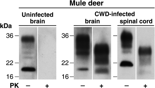
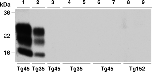
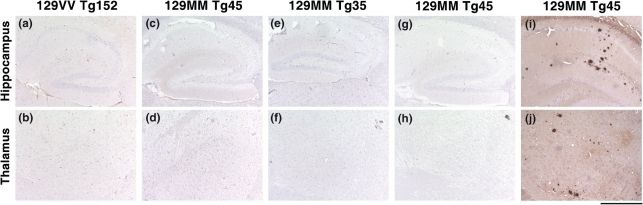
Chronic wasting disease (CWD) is a prion disease that affects free-ranging and captive cervids, including mule deer, white-tailed deer, Rocky Mountain elk and moose. CWD-infected cervids have been reported in 14 USA states, two Canadian provinces and in South Korea. The possibility of a zoonotic transmission of CWD prions via diet is of particular concern in North America where hunting of cervids is a popular sport. To investigate the potential public health risks posed by CWD prions, we have investigated whether intracerebral inoculation of brain and spinal cord from CWD-infected mule deer transmits prion infection to transgenic mice overexpressing human prion protein with methionine or valine at polymorphic residue 129. These transgenic mice have been utilized in extensive transmission studies of human and animal prion disease and are susceptible to BSE and vCJD prions, allowing comparison with CWD. Here, we show that these mice proved entirely resistant to infection with mule deer CWD prions arguing that the transmission barrier associated with this prion strain/host combination is greater than that observed with classical BSE prions. However, it is possible that CWD may be caused by multiple prion strains. Further studies will be required to evaluate the transmission properties of distinct cervid prion strains as they are characterized.
Figures
References
-
- Anderson, C. A., Bosque, P., Filley, C. M., Arciniegas, D. B., Kleinschmidt-Demasters, B. K., Pape, W. J. & Tyler, K. L. (2007). Colorado surveillance program for chronic wasting disease transmission to humans: lessons from 2 highly suspicious but negative cases. Arch Neurol 64, 439–441. - PubMed
-
- Angers, R. C., Browning, S. R., Seward, T. S., Sigurdson, C. J., Miller, M. W., Hoover, E. A. & Telling, G. C. (2006). Prions in skeletal muscles of deer with chronic wasting disease. Science 311, 1117. - PubMed
-
- Asante, E. A., Linehan, J. M., Desbruslais, M., Joiner, S., Gowland, I., Wood, A. L., Welch, J., Hill, A. F., Lloyd, S. E. & other authors (2002). BSE prions propagate as either variant CJD-like or sporadic CJD-like prion strains in transgenic mice expressing human prion protein. EMBO J 21, 6358–6366. - PMC - PubMed
-
- Asante, E. A., Linehan, J. M., Gowland, I., Joiner, S., Fox, K., Cooper, S., Osiguwa, O., Gorry, M., Welch, J. & other authors (2006). Dissociation of pathological and molecular phenotype of variant Creutzfeldt–Jakob disease in transgenic human prion protein 129 heterozygous mice. Proc Natl Acad Sci U S A 103, 10759–10764. - PMC - PubMed
Publication types
MeSH terms
Substances
Grants and funding
LinkOut - more resources
Full Text Sources