. 2010 Sep;84(18):9533-45.
doi: 10.1128/JVI.01005-10. Epub 2010 Jul 7.

Role of peroxiredoxin 1 and peroxiredoxin 4 in protection of respiratory syncytial virus-induced cysteinyl oxidation of nuclear cytoskeletal proteins

Affiliations

Role of peroxiredoxin 1 and peroxiredoxin 4 in protection of respiratory syncytial virus-induced cysteinyl oxidation of nuclear cytoskeletal proteins

Mohammad Jamaluddin et al. J Virol. 2010 Sep.

Abstract

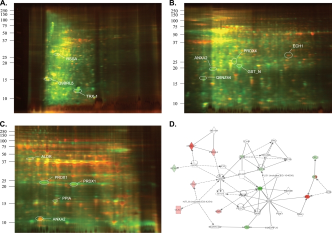
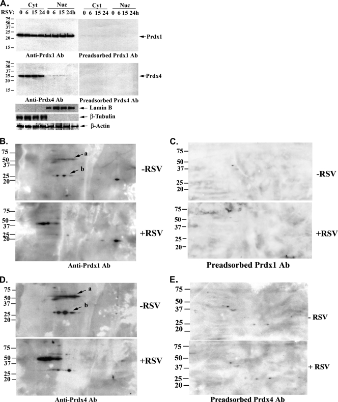
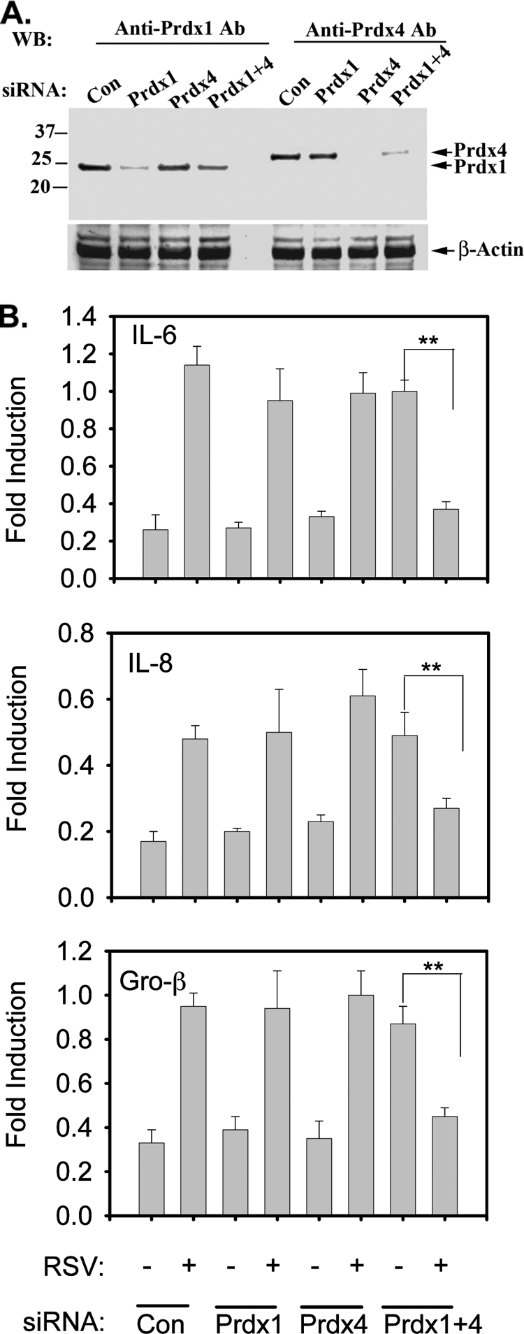
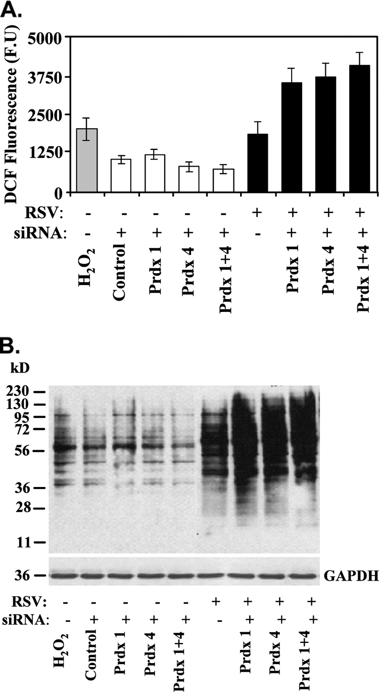
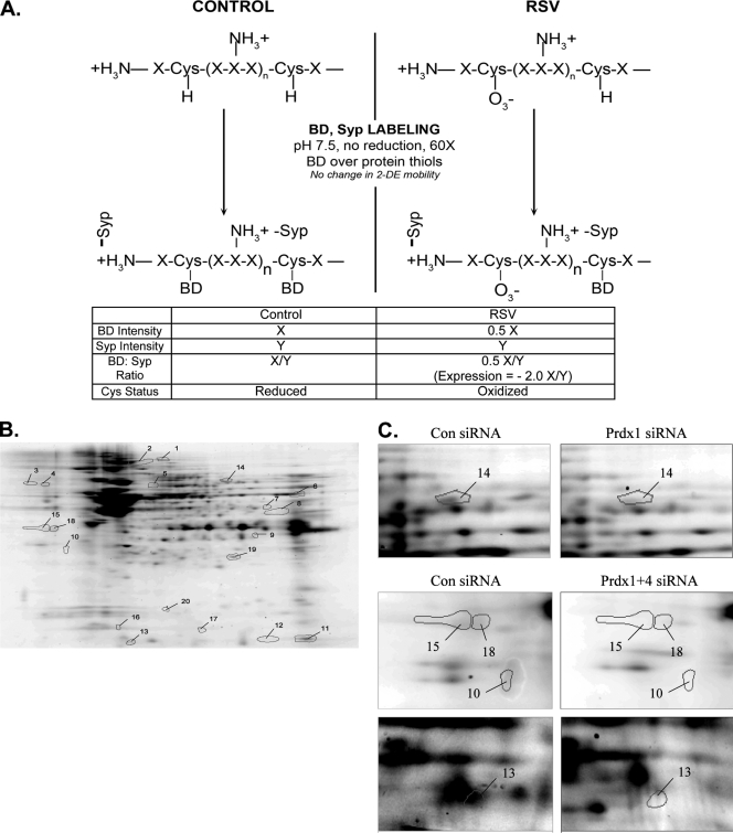
The respiratory epithelium plays a central role in innate immunity by secreting networks of inflammatory mediators in response to respiratory syncytial virus (RSV) infection. Previous proteomic studies focusing on the host cellular response to RSV indicated the existence of a nuclear heat shock response and cytoplasmic depletion of antioxidant proteins in model type II-like airway epithelial cells. Here, we increased the depth of nuclear proteomic interrogation by using fluorescence difference labeling followed by liquid isoelectric focusing prefractionation/two-dimensional gel electrophoresis (2-DE) to identify an additional 41 proteins affected by RSV infection. Surprisingly, we found inducible oligomers and shifts in isoelectric points for peroxiredoxin 1 (Prdx-1), Prdx-3, and Prdx-4 isoforms without changes in their total abundance, indicating that Prdxs were being oxidized in response to RSV. To address the role of Prdx-1 and Prdx-4 in RSV infection, isoforms were selectively knocked down by small interfering RNA (siRNA) transfection. Cells lacking Prdx-1, Prdx-4, or both showed increased levels of reactive oxygen species formation and a higher level of protein carbonylation in response to RSV infection. Using a novel saturation fluorescence labeling 2-DE analysis, we showed that 15 unique proteins had enhanced oxidative modifications of at least >1.2-fold in the Prdx knockdowns in response to RSV, including annexin A2 and desmoplakin. Our results suggest that Prdx-1 and Prdx-4 are essential for preventing RSV-induced oxidative damage in a subset of nuclear intermediate filament and actin binding proteins in epithelial cells.


Figures

FIG. 1.
FIG. 1.
RSV-induced changes of nuclear proteins of A549 cells by 2-D DIGE. Dual channel overlay of representative gel images in the pH ranges of 3.0 to 5.4 (A), 5.4 to 7.0 (B), and 7.0 to 10.0 (C). Uninfected and RSV-infected nuclear extracts were labeled with Cy3 (green) and Cy5 (red), respectively. For clarity, only a few representative spots are illustrated and found in Table 1. (D) Graphical representation of a pathway containing the largest number of focus proteins. Each node represents individual proteins, and relationships are represented by lines. Nodes colored red indicate a protein differentially upregulated by RSV, while nodes colored green indicate a downregulated protein. Uncolored nodes are inferred. Squares indicate cytokines, circles indicate chemokines, and ovals indicate transcription factors. Lines terminating with an arrow indicate “acts on.” Act6A, actin binding protein; CFL, cofilin; ECH, enoyl coenzyme A hydratase; KRT, keratin; Jnk, Jun N-terminal kinase; Prdx, peroxiredoxin; VMT, vimentin.
FIG. 2.
FIG. 2.
Validation of peroxiredoxin (Prdx) 1 and Prdx-4 expression in A549 cells by Western immunoblotting. (A) Effect of RSV infection on cytoplasmic-nuclear localization of Prdx-1 and Prdx-4 in A549 cells by Western blotting. Cells were infected with RSV for 0 to 24 h, and 50 μg of cytoplasmic (Cyt) or nuclear (Nuc) extracts were separated on a 12% SDS-PAGE gel and transferred to PVDF membranes. Membranes were probed with either Prdx-1 antibody (anti-Prdx-1 Ab) or Prdx-4 Ab (anti-Prdx-4 Ab) (left) or immunogen peptide-preadsorbed Prdx-1 and Prdx-4 Abs (right). After the membranes were stripped, the membranes were probed with either anti-Lam B, anti-β-tubulin, or anti-β-actin Abs. Molecular weight markers are shown on the left. (B) Induction of Prdx-1 by RSV infection. Nuclear extracts (150 μg) from uninfected (−RSV) or RSV-infected (+RSV) A549 cells were separated on a 2-D gel and transferred to PVDF membranes. Membranes were probed with Prdx-1 Ab (anti-Prdx-1 Ab). Arrows labeled “a” and “b” indicate dimeric and monomeric forms of Prdx-1, respectively. Molecular weight markers are shown on the left. (C) Prdx-1 Ab specificity. Identical amounts of proteins were separated by 2-DE and transferred to Immobilon membranes. Prdx-1 Ab was preadsorbed by mixing with blocking peptides before exposure to membranes. (D) RSV-induced changes of Prdx-4 by 2-DE Western blotting. Prdx-4 was detected by Western blotting using Prdx-4 Ab (anti-Prdx-4 Ab). The arrow labeled “a” designates dimer; the arrow labeled “b” designates the monomer of Prdx-4. (E) Prdx-4 Ab was preadsorbed prior to blotting, as described for panel C.
FIG. 3.
FIG. 3.
siRNA-mediated knockdown of Prdx-1 and Prdx-4 in RSV-treated A549 cells. Cells were transfected with 100 nM scrambled (Con), Prdx-1, Prdx-4, or Prdx-1 plus Prdx-4 siRNA. (A) After 72 h, cells were harvested and Prdx-1 and Prdx-4 were detected in whole-cell extract (WCE) by Western immunoblotting. Τhe same membrane was probed with β-actin Ab as a loading control. (B) A549 cells were transfected with either scrambled, Prdx-1, Prdx-4, or Prdx-1 plus Prdx-4 siRNA for 72 h, followed by no infection or RSV infection for 15 h. Total RNA was extracted and subjected to quantitative reverse transcription-PCR (qRT-PCR) for IL-6, Gro-β, and IL-8. Data are normalized to glyceraldehyde-3-phosphate dehydrogenase (GAPDH) and expressed as fold changes relative to the uninfected control. Each bar is the mean ± standard deviation from triplicate determinations. **, P < 0.001.
FIG. 4.
FIG. 4.
ROS production in A549 cells lacking Prdx-1, Prdx-4, or both. (A) ROS generation was measured by monitoring changes in oxidized DCF fluorescence in A549 cells transfected with control, Prdx-1, Prdx-4, or both Prdx-1 and Prdx-4 siRNA, after which the cells were infected with RSV for 15 h. Bars represent the means ± standard errors of two independent measurements in duplicate. H2O2 was used as a positive control. (B) Enhanced carbonylation of proteins by RSV infection in Prdx-1, Prdx-4, or Prdx-1 plus Prdx-4 knockdown cells by Western blotting. Cellular extracts from an experiment identical to that described for panel A were subjected to SDS-PAGE and transferred to a PVDF membrane. The membrane was probed with anti-DNP antibody. GAPDH is shown as a loading control.
FIG. 5.
FIG. 5.
Saturation fluorescence labeling with BODIPY-FL. (A) Schematic diagram of an example of the saturation fluorescence labeling assay for detection of cysteine oxidation. Protein extracts were assayed for cysteine content by amino acid analysis. Enough BODIPY-FL-maleimide was added at pH 7.5 to the unreduced protein mixture to yield a ratio of 60-fold BODIPY to protein thiols. BODIPY will react with reduced cysteine thiols but will not label oxidized thiols. Sypro ruby stain is directed to free amino groups and not affected by cysteine oxidation. The BODIPY/Sypro ruby ratio reflects the oxidation status of cysteine relative to the total protein. The example shows only the partial oxidation of the protein; complete cysteinyl oxidation would result in a very low BODIPY/Sypro ratio, indicating extreme oxidation. BD, BODIPY; Syp, Sypro ruby. (B) A representative image of a 2-D gel of proteins labeled with BODIPY. Nuclear extracts from A549 cells transfected with Prdx-1 siRNA and infected with RSV were labeled with BODIPY and separated by 2-DE. The numbers on the gel indicate individual protein spots whose abundance has changed >1.2-fold. These were identified by electrospray tandem MS on the OrbiTrap Velo. Their identities are listed in Table 2. (C) Expanded view of a few exemplary nuclear protein spots with notable differences in abundances from cells transfected with control, Prdx-1, or Prdx-1 plus Prdx-4 siRNAs.
FIG. 6.
FIG. 6.
Network analysis of nuclear proteins which have been oxidized by RSV infection in Prdx-1- or Prdx-4-silenced A549 cells. Shown is the molecular interaction network program using the 15 unique proteins found to be most oxidized by RSV infection as the input. Note that all 15 unique proteins occur in the network (gray). ALDH1B1, aldehyde dehydrogenase 1 family, member B1; ANXA2, annexin A2; ATF4, activating transcription factor 4 (tax-responsive enhancer element B67); ATP5A1, ATP synthase, H+ transporting, mitochondrial F1 complex, alpha subunit 1, cardiac muscle; CDC42SE1, CDC42 small effector 1; CENPE, centromere protein E, 312 kDa; CEP290, centrosomal protein, 290 kDa; CPS1, carbamoyl-phosphate synthetase 1, mitochondrial; DSP, desmoplakin; ERBB2, v-erb-b2 erythroblastic leukemia viral oncogene homolog 2, neuro/glioblastoma-derived oncogene homolog (avian); GFAP, glial fibrillary acidic protein; GOT2, glutamic-oxaloacetic transaminase 2, mitochondrial (aspartate aminotransferase 2); HADHA, hydroxyacyl-coenzyme A dehydrogenase/3-ketoacyl-coenzyme A thiolase/enoyl-coenzyme A hydratase (trifunctional protein), alpha subunit; HSPA9, heat shock 70-kDa protein 9 (mortalin); IMPACT, impact homolog (mouse); LEP, leptin; LMNA, Lam A/C; LRPPRC, leucine-rich PPR-motif containing; MAPK8, mitogen-activated protein kinase 8; MYC, v-myc myelocytomatosis viral oncogene homolog (avian); NFKB2, nuclear factor of kappa light polypeptide gene enhancer in B cells 2 (p49/p100); NNMT, nicotinamide N-methyltransferase; PAK1IP1, PAK1 interacting protein 1; PPL, periplakin; Prdx5, peroxiredoxin 5; PRL2C3, prolactin family 2, subfamily c, member 3; RPL17, ribosomal protein L17; SLC11A1, solute carrier family 11 (proton-coupled divalent metal ion transporters), member 1; SOLH, small optic lobes homolog (Drosophila); SP1, Sp1 transcription factor; SPAG4, sperm-associated antigen 4; SQRDL, sulfide quinone reductase-like (yeast); STMN3, stathmin-like 3; TGFB1, transforming growth factor, beta 1.

References

    1. Aguilera-Aguirre, L., A. Bacsi, A. Saavedra-Molina, A. Kurosky, S. Sur, and I. Boldogh. 2009. Mitochondrial dysfunction increases allergic airway inflammation. J. Immunol. 183:5379-5387. - PMC - PubMed
    1. Boldogh, I., A. Basci, B. Choudhary, N. Dharajiya, R. Alam, T. Hazdra, S. Mitra, R. Goldblum, and S. Sur. 2005. ROS generated by pollen NADPH oxidase provide a signal that augments antigen-induced allergic airway inflammation. J. Clin. Invest. 115:2169-2179. - PMC - PubMed
    1. Brasier, A. R., H. Spratt, Z. Wu, I. Boldogh, Y. Zhang, R. P. Garofalo, A. Casola, J. Pashmi, A. Haag, B. Luxon, and A. Kurosky. 2004. Nuclear heat shock response and novel nuclear domain 10 reorganization in respiratory syncytial virus-infected A549 cells identified by high-resolution two-dimensional gel electrophoresis. J. Virol. 78:11461-11476. - PMC - PubMed
    1. Caplan, J. F., N. R. Filipenko, S. L. Fitzpatrick, and D. M. Waisman. 2004. Regulation of annexin A2 by reversible glutathionylation. J. Biol. Chem. 279:7740-7750. - PubMed
    1. Casola, A., N. Burger, T. Liu, M. Jamaluddin, A. R. Brasier, and R. P. Garofalo. 2001. Oxidant tone regulates RANTES gene expression in airway epithelial cells infected with respiratory syncytial virus. Role in viral-induced interferon regulatory factor activation. J. Biol. Chem. 276:19715-19722. - PubMed

Publication types