. 2010 Sep;84(18):9318-25.
doi: 10.1128/JVI.01049-10. Epub 2010 Jul 7.

T cell responses are required for protection from clinical disease and for virus clearance in severe acute respiratory syndrome coronavirus-infected mice

Affiliations

T cell responses are required for protection from clinical disease and for virus clearance in severe acute respiratory syndrome coronavirus-infected mice

Jincun Zhao et al. J Virol. 2010 Sep.

Abstract

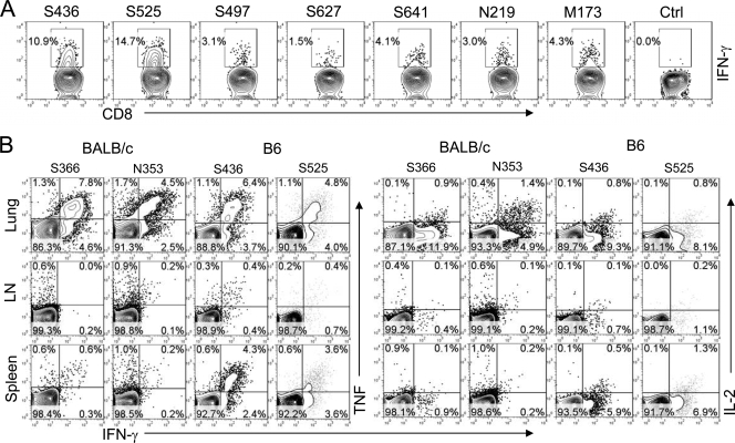
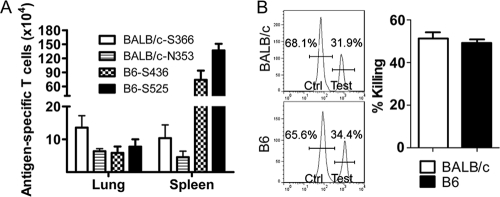
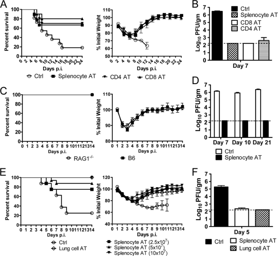
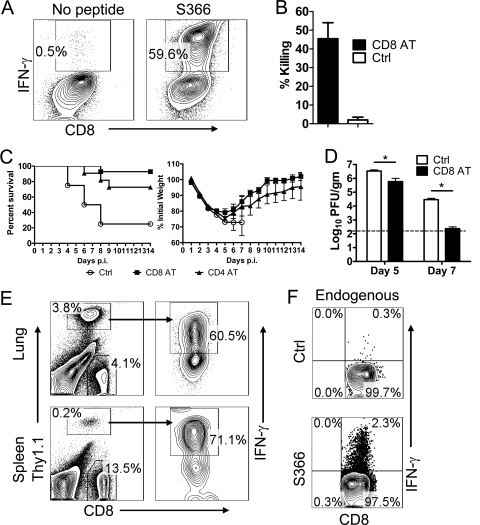
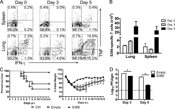
A dysregulated innate immune response and exuberant cytokine/chemokine expression are believed to be critical factors in the pathogenesis of severe acute respiratory syndrome (SARS), caused by a coronavirus (SARS-CoV). However, we recently showed that inefficient immune activation and a poor virus-specific T cell response underlie severe disease in SARS-CoV-infected mice. Here, we extend these results to show that virus-specific T cells, in the absence of activation of the innate immune response, were sufficient to significantly enhance survival and diminish clinical disease. We demonstrated that T cells are responsible for virus clearance, as intravenous adoptive transfer of SARS-CoV-immune splenocytes or in vitro-generated T cells to SCID or BALB/c mice enhanced survival and reduced virus titers in the lung. Enhancement of the number of virus-specific CD8 T cells by immunization with SARS-CoV peptide-pulsed dendritic cells also resulted in a robust T cell response, earlier virus clearance, and increased survival. These studies are the first to show that T cells play a crucial role in SARS-CoV clearance and that a suboptimal T cell response contributes to the pathological changes observed in SARS. They also provide a new approach to SARS vaccine design.


Figures

FIG. 1.
FIG. 1.
T cell epitope identification and cytokine production in MA15 virus-infected BALB/c and B6 mice. (A) B6 mice (6 to 8 weeks old) were infected intranasally with 3 × 10⁵ PFU MA15 virus in 50 μl DMEM. BALB/c mice were treated with clodronate, because in its absence, most BALB/c mice do not mount a virus-specific T cell response after infection with this virus dosage (35). At day 8, single cell suspensions were prepared from lungs and stimulated with 1 μM SARS-CoV-specific CD8 or CD4 T cell peptides for 5 h in the presence of brefeldin A. Frequencies of MA15 virus-specific T cells (determined by IFN-γ intracellular staining) are shown. Data are representative of three independent experiments, with 5 to 8 mice/group. (B) BALB/c or B6 mice (6 to 8 weeks old) were infected intranasally with 3 × 10³ PFU or 3 × 10⁵ PFU MA15 virus, respectively, in 50 μl DMEM. At day 7, single cell suspensions were prepared from lungs, LNs, and spleens and stimulated with SARS-CoV CD8 or CD4 T cell peptides for 5 h in the presence of brefeldin A. IL-2, TNF-α, and IFN-γ expression levels are shown. Data are summarized in Fig. 2A.
FIG. 2.
FIG. 2.
Characterization of T cell response in MA15 virus-infected BALB/c and B6 mice. (A) Numbers of MA15 virus-specific T cells are shown. Data are representative of two independent experiments, with 4 mice/group. (B) In vivo cytotoxicity assays were performed on day 6 p.i. as described in Materials and Methods. Data are representative of three independent experiments, with 3 or 4 mice/group.
FIG. 3.
FIG. 3.
SARS-CoV-specific T cell transfer cleared virus and ameliorated clinical disease in both immunocompromised and immunocompetent mice. Donor BALB/c mice were treated with clodronate 1 day prior to infection with 3 × 10⁴ PFU MA15 virus. Donor B6 mice were infected with 3 × 10⁵ PFU MA15. Mice were sacrificed 8 days p.i. and cells harvested from the infected lungs and spleens. Recipient SCID (A and B), B6 and RAG1−/− (C and D), or BALB/c (E and F) mice (6 to 8 weeks old) received 2.5 × 10⁷, 5 × 10⁷, or 10 × 107 splenocytes, 2 × 10⁷ total lung cells, or 5 × 10⁶ lung-derived CD4 or CD8 T cells at day −1 or no cells and were then infected intranasally with 3 × 10⁴ PFU (SCID and BALB/c mice) or 3 × 10⁵ PFU (B6 and RAG1−/− mice) MA15 virus in 50 μl DMEM at day 0. Mortality and weight were monitored daily. (A) Recipient SCID mice. Eleven mice were in the control group, 9 mice received 5 × 10⁷ total splenocytes, and 10 mice received lung-derived CD4 or CD8 T cells. Survival was significantly greater in recipients of CD4 (P < 0.05) or CD8 (P < 0.01) T cell transfer and nearly significantly greater after total splenocyte transfer (P = 0.06). (C) B6 and RAG1−/− mice. Eight mice were in each group. B6 and RAG1−/− mice showed mild weight loss but remained asymptomatic after MA15 virus infection even in the absence of adoptively transferred cells. (E) BALB/c mice. Eight mice were in the control group; 8, 16, or 8 mice received 2.5 × 10⁷, 5 × 10⁷, or 10 × 107 splenocytes, respectively; and 8 mice received total lung cells. Survival was significantly greater after transfer of 5 × 10⁷ or 10 × 107 splenocytes (P < 0.007 for both) or lung cells (P < 0.007). For virus titers, infected SCID (B), RAG1−/− (D), or BALB/c (F) mice received adoptively transferred cells or no cells and were sacrificed at the indicated time points. Lung homogenates were prepared, and titers were determined on Vero E6 cells. Viral titers are expressed as PFU/g tissue. There were 4 mice/group. AT, adoptive transfer. The horizontal line denotes the limit of detection.
FIG. 4.
FIG. 4.
Transfer of SARS-CoV-specific CD8 T cell lines decreased virus titers and ameliorated clinical disease in BALB/c mice. Epitope S366-specific CD8 or epitope N353-specific CD4 T cell lines (TCLs) were propagated in vitro as described in Materials and Methods. (A) TCLs were stimulated with SARS-CoV CD8 peptide for 5 h in the presence of brefeldin A and examined for expression of IFN-γ. (B) In vivo cytotoxicity assays were performed 1 day after transfer of CD8 T cells as described in Materials and Methods. Data are representative of two independent experiments, with 3 or 4 mice/group. (C) CD8 or CD4 T cells (1 × 10⁷) were transferred to 6- to 8-week-old BALB/c mice 1 day prior to i.n. infection with 3 × 10⁴ PFU MA15 virus. Mortality was monitored daily. Eight mice were in the control group, 14 mice received CD8 T cells (P = 0.002), and 11 mice received CD4 T cells (P = 0.07). (D) For virus titers, lungs were homogenized at the indicated time points and titers were determined on Vero E6 cells. Viral titers are expressed as PFU/g tissue. There were 4 mice/group. The horizontal line indicates the limit of detection. *, P < 0.05. (E) Single cell suspensions were prepared from the lungs and spleens of infected BALB/c mice that had received 1 × 10⁷ CD8 T cells. Cells were stimulated with SARS-CoV-specific CD8 T cell peptide S366 for 5 h in the presence of brefeldin A. IFN-γ expression frequencies of transferred T cells are shown. (F) Endogenous CD8 T cell responses at day 6 p.i. in recipients of transferred CD8 T cells were determined by IFN-γ intracellular staining. Data are representative of two independent experiments, with 4 mice/group.
FIG. 5.
FIG. 5.
Immunization with DC/peptide S366 complexes induced protective CD8 T cell responses. BALB/c mice (6 to 8 weeks old) were immunized with BMDCs pulsed with peptide S366. Seven days later, mice were infected intranasally with 3 × 10⁴ PFU MA15 virus. Single cell suspensions were prepared from lungs and spleens at the indicated time points and stimulated with SARS-CoV S366 peptide for 5 h in the presence of brefeldin A. TNF-α and IFN-γ expression frequencies (A) and numbers (B) of SARS-CoV-specific T cells are shown. (C) Mortality was monitored daily. Ten mice were in the control group, and 10 and 19 mice were immunized with uncoated BMDCs (empty) and peptide S366-coated BMDCs, respectively. Survival was significantly greater in recipients of S366-coated BMDCs than in those receiving no (control) or empty BMDCs (P < 0.02). (D) For virus titers, lungs were homogenized at the indicated time points and titers were determined on Vero E6 cells. Viral titers are expressed as PFU/g tissue. There were 4 mice/group. The horizontal line denotes the limit of detection.

References

    1. Baas, T., A. Roberts, T. H. Teal, L. Vogel, J. Chen, T. M. Tumpey, M. G. Katze, and K. Subbarao. 2008. Genomic analysis reveals age-dependent innate immune responses to severe acute respiratory syndrome coronavirus. J. Virol. 82:9465-9476. - PMC - PubMed
    1. Badovinac, V. P., K. A. Messingham, A. Jabbari, J. S. Haring, and J. T. Harty. 2005. Accelerated CD8+ T-cell memory and prime-boost response after dendritic-cell vaccination. Nat. Med. 11:748-756. - PubMed
    1. Barber, D. L., E. J. Wherry, and R. Ahmed. 2003. Cutting edge: rapid in vivo killing by memory CD8 T cells. J. Immunol. 171:27-31. - PubMed
    1. Cameron, M. J., L. Ran, L. Xu, A. Danesh, J. F. Bermejo-Martin, C. M. Cameron, M. P. Muller, W. L. Gold, S. E. Richardson, S. M. Poutanen, B. M. Willey, M. E. DeVries, Y. Fang, C. Seneviratne, S. E. Bosinger, D. Persad, P. Wilkinson, L. D. Greller, R. Somogyi, A. Humar, S. Keshavjee, M. Louie, M. B. Loeb, J. Brunton, A. J. McGeer, S. R. N. Canadian, and D. J. Kelvin. 2007. Interferon-mediated immunopathological events are associated with atypical innate and adaptive immune responses in patients with severe acute respiratory syndrome. J. Virol. 81:8692-8706. - PMC - PubMed
    1. Chen, J., Y. F. Lau, E. W. Lamirande, C. D. Paddock, J. H. Bartlett, S. R. Zaki, and K. Subbarao. 2010. Cellular immune responses to severe acute respiratory syndrome coronavirus (SARS-CoV) infection in senescent BALB/c mice: CD4+ T cells are important in control of SARS-CoV infection. J. Virol. 84:1289-1301. - PMC - PubMed

Publication types

MeSH terms