. 2010 Jul 7;30(27):9027-37.
doi: 10.1523/JNEUROSCI.1635-10.2010.

Interplay of palmitoylation and phosphorylation in the trafficking and localization of phosphodiesterase 10A: implications for the treatment of schizophrenia

Affiliations

Interplay of palmitoylation and phosphorylation in the trafficking and localization of phosphodiesterase 10A: implications for the treatment of schizophrenia

Erik I Charych et al. J Neurosci. .

Abstract

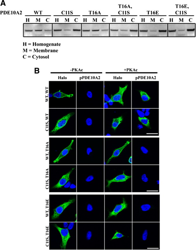
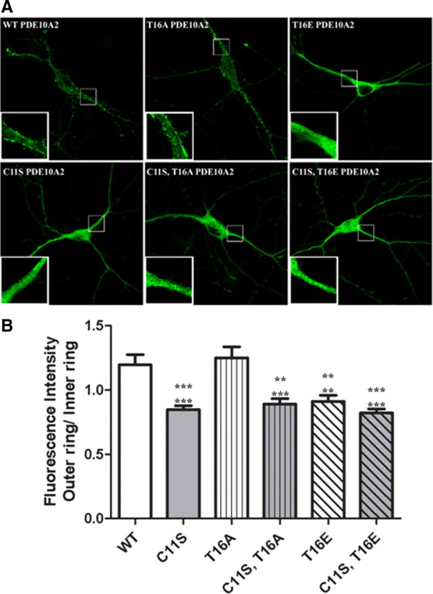
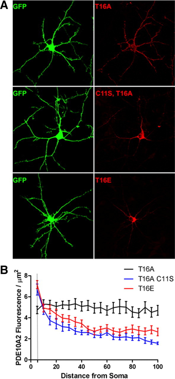
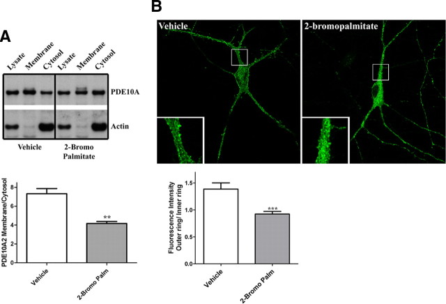
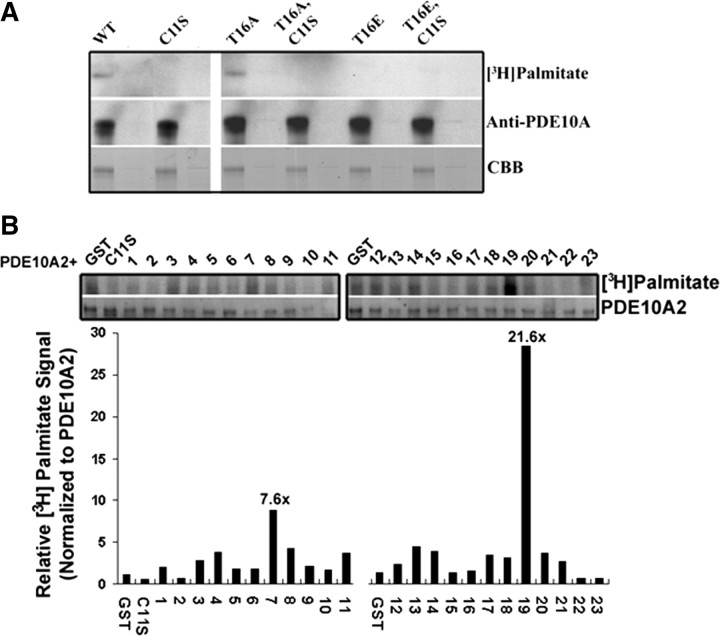
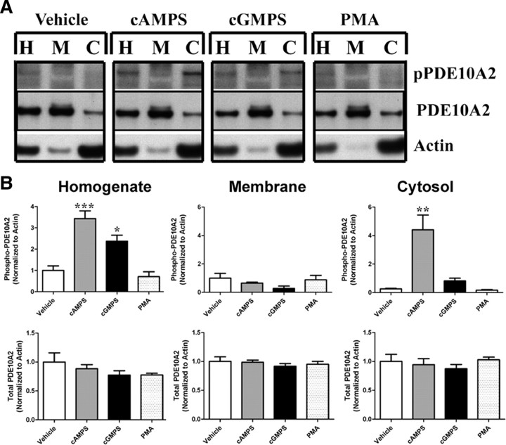
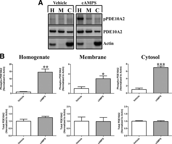
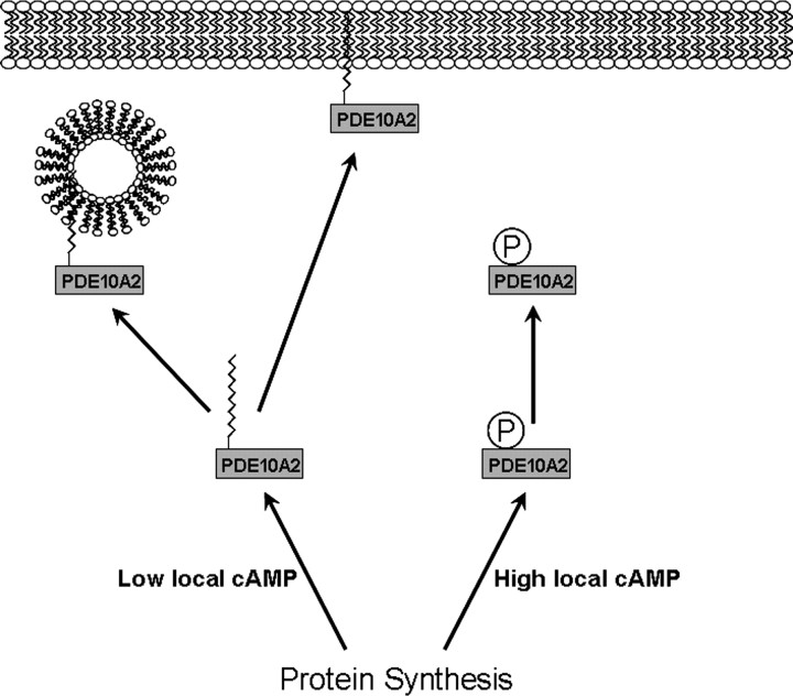
Phosphodiesterase 10A (PDE10A) is a striatum-enriched, dual-specific cyclic nucleotide phosphodiesterase that has gained considerable attention as a potential therapeutic target for psychiatric disorders such as schizophrenia. As such, a PDE10A-selective inhibitor compound, MP-10, has recently entered clinical testing. Since little is known about the cellular regulation of PDE10A, we sought to elucidate the mechanisms that govern its subcellular localization in striatal medium spiny neurons. Previous reports suggest that PDE10A is primarily membrane bound and is transported throughout medium spiny neuron axons and dendrites. Moreover, it has been shown in PC12 cells that the localization of the major splice form, PDE10A2, may be regulated by protein kinase A phosphorylation at threonine 16 (Thr-16). Using an antibody that specifically recognizes phosphorylated Thr-16 (pThr-16) of PDE10A2, we provide evidence that phosphorylation at Thr-16 is critical for the regulation of PDE10A subcellular localization in vivo. Furthermore, we demonstrate in primary mouse striatal neuron cultures that PDE10A membrane association and transport throughout dendritic processes requires palmitoylation of cysteine 11 (Cys-11) of PDE10A2, likely by the palmitoyl acyltransferases DHHC-7 and -19. Finally, we show that Thr-16 phosphorylation regulates PDE10A trafficking and localization by preventing palmitoylation of Cys-11 rather than by interfering with palmitate-lipid interactions. These data support a model whereby PDE10A trafficking and localization can be regulated in response to local fluctuations in cAMP levels. Given this, we propose that excessive striatal dopamine release, as occurs in schizophrenia, might exert differential effects on the regulation of PDE10A localization in the two striatal output pathways.


Figures

Figure 1.
Figure 1.
Phosphorylation at Thr-16 leads to a cytosolic localization of endogenous PDE10A2 in mouse striatum. A, The difference in localization pattern between the two major PDE10A splice variants in rodent and human, PDE10A1 and PDE10A2, is caused by alternative splicing in the region encoding the extreme N terminus of PDE10A. B, Anti-pPDE10A2 strongly reacted with a bacterially expressed N-terminal fragment of PDE10A2 only when this fragment was phosphorylated in vitro by PKA and contained a threonine, but not an alanine, at position 16. The immunoreactive band was displaced by a phosphorylated, but not an unphosphorylated, immunogenic peptide. C, When coexpressed with PKAc in transfected HEK293 cells, both PDE10A2WT and PDE10A2T16E, the latter carrying the phosphomimetic mutation at amino acid position 16, were enriched in the cytosolic (Cyt) fraction over the membrane (Mem) fraction (top), but only PDE10A2WT was phosphorylated at Thr-16 (bottom). In contrast, PDE10A2T16A, which is predicted to be resistant to phosphorylation at Thr-16 by PKAc, is enriched in the membrane fraction over the cytosolic fraction (top) and is not phosphorylated by PKAc (bottom). D, Phospho-PDE10A2 was detected in immunoprecipitates (IP) of total PDE10A in mouse striatal extracts. The anti-pPDE10A2 immunoreactive band was displaced by the phosphorylated, but not the nonphosphorylated, immunogenic peptide. The signal strength of the anti-pPDE10A2 band was further elevated by in vitro phosphorylation by PKAc. E, Crude membrane and cytosolic fractions of mouse striatum were immunoblotted with anti-pPDE10A2 or anti-PDE10A. The ratio of immunoreactive band intensities in the cytosolic versus membrane fractions are shown for both anti-pPDE10A2 and anti-PDE10A. Phosphorylated PDE10A2 is highly enriched in the cytosolic fraction over the membrane fraction of the mouse striatum. Conversely, total PDE10A is enriched in the membrane fraction over the cytosolic fraction of mouse striatum. ***p < 0.001 by Student's t test.
Figure 2.
Figure 2.
Plasma membrane binding of PDE10A2 requires Cys-11 in HEK293 cells. A, A mutant form of PDE10A2 containing a serine in place of Cys-11 (PDE10A2C11S) was enriched in the cytosol as determined by Western blot analysis of crude membrane and cytosolic fractions of HEK293 cells transfected with the indicated PDE10A2 mutants. B, Immunofluorescence confocal microscopy using antibodies to total and phosphorylated PDE10A2 showed that the C11S mutation abolished plasma membrane binding independent of the amino acid at position 16 and of the phosphorylation state of PDE10A2, as determined by pPDE10A2 immunofluorescence labeling. Halo, HaloTag antibody.
Figure 3.
Figure 3.
Cys-11 is required for association of PDE10A2 to the cytoplasmic face of the plasma membrane in cultured striatal neurons. A, Both PDE10A2WT and PDE10A2T16A immunofluorescence was strongly accumulated in the plasma membrane in the somata and dendrites (insets) of transfected primary striatal neurons. In contrast, PDE10A2T16E, PDE10A2C11S, PDE10A2C11S/T16A, and PDE10A2C11S/T16E immunofluorescence was strongly accumulated in the cytosol, both in the soma and in dendrites (insets) of these cells. B, Quantification of the degree of membrane enrichment of immunofluorescent labeling over the cytosol (see Materials and Methods for details) in transfected striatal neuron cultures. The outer ring (membrane immunofluorescence) to inner ring (cytosolic immunofluorescence) ratio was significantly greater for both PDE10A2WT and PDE10A2T16A compared with PDE10A2T16E, PDE10A2C11S, PDE10A2C11S/T16A, or PDE10A2C11S/T16E. PDE10A2T16D and PDE10A2C11S/T16D (data not shown) also showed significant reductions in outer ring to inner ring ratio compared with both PDE10A2WT and PDE10A2T16A. ***p < 0.001; **p < 0.01 by one-way ANOVA followed by Dunnett's multiple comparison test. Upper asterisks denote comparison with PDE10A2WT; lower asterisks denote comparison with PDE10A2T16A. No significant difference between PDE10A2WT and PDE10A2T16A was observed.
Figure 4.
Figure 4.
Distal dendritic trafficking of PDE10A2 requires Cys-11 in cultured striatal neurons. A, Representative primary cultures of striatal neurons transfected with cDNA encoding EGFP and either the phospho-resistant PDE10A2T16A, the phosphomimetic PDE10A2T16E, or the membrane binding-deficient PDE10A2C11S mutant. B, Confocal images were analyzed by generating 5 × 3 μm bins at identical positions in both the EGFP channel and the PDE10A2 immunofluorescence channel that extended sequentially for 100 μm along dendritic arbors beginning at the soma boundaries. For each bin, total area occupied by EGFP and the average PDE10A2 fluorescence intensity was measured. The PDE10A2 fluorescence normalized to dendritic area was plotted against the distance of that segment from the soma boundary. Fluorescence/μm2 for the phospho-resistant PDE10A2T16A mutant, which contained an intact cysteine at position 11, did not differ as function of distance from the soma. In contrast, fluorescence/μm2 of the phosphomimetic PDE10A2T16E mutant or the membrane binding-deficient PDE10A2C11S mutant was greatest near the soma boundary and sharply decayed with increasing distance from the soma boundary. Two-way repeated-measures ANOVA, effect of distance × treatment, p < 0.0001. Post hoc analysis by least significant difference: PDE10A2T16A, 0 μm versus any other distance, NS; PDE10A2C11S, 0 μm versus 5–100 μm, p < 0.0001 for each comparison; PDE10A2T16E, 0 μm versus 5–100 μm, p < 0.0001 for each comparison.
Figure 5.
Figure 5.
PDE10A2 membrane binding is disrupted by inhibiting palmitoyltransferase activity. A, Crude fractionation of HEK293 cells expressing the phospho-resistant PDE10A2T16A mutant showed that the PDE10A2 membrane to cytosol ratio was significantly reduced after treatment with 100 μm 2-bromopalmitate compared with vehicle control. B, The ratio of outer ring to inner ring fluorescence intensity was significantly reduced after primary striatal neurons, transfected with the phospho-resistant PDE10A2T16A mutant, were treated with 100 μm 2-bromopalmitate. **p < 0.01; ***p < 0.001 by Student's t test.
Figure 6.
Figure 6.
PDE10A2 is palmitoylated on Cys-11. A, Palmitoylation assays show that PDE10A2WT and PDE10A2T16A incorporated [3H]palmitate, whereas PDE10A2C11S and PDE10A2T16E did not, suggesting that Cys-11 is necessary for palmitoylation of PDE10A2, but Thr-16 must also be unphosphorylated. B, To identify the PATs likely to mediate the palmitoylation of native PDE10A2, HEK293 cells were transfected with the 23 cloned mouse ZDHHC proteins and PDE10A2 (WT or C11S) using the [3H]palmitate incorporation assay. As shown, ZDHHC-7 and -19 elicited robust [3H]palmitate signals over basal levels.
Figure 7.
Figure 7.
Acute phosphorylation of Thr-16 does not cause translocation of PDE10A2. A, HEK293 cells expressing PDE10A2 were treated for 1 h with a membrane-permeant, PDE-resistant cAMP analog (Sp-6-Phe-cAMPS), a membrane-permeant, PDE-resistant cGMP analog (Sp-8-pCPT-cGMPS), or PMA, and crude fractions were immunoblotted with anti-pPDE10A2 and anti-PDE10A to test whether acute phosphorylation of Thr-16 leads to a disengagement of PDE10A2 with cellular membranes. H, Homogenate, M, membrane, C, cytosol. B, Treatment with Sp-6-Phe-cAMPS lead to a robust increase in phosphorylation of PDE10A2 at Thr-16 in total homogenates, as well as in the cytosolic fraction. Treatment with Sp-8-pCPT-cGMPS elicited a significant increase in phosphorylation of PDE10A2 at Thr-16 in total homogenates only. PMA did not alter the phosphorylation state of PDE10A2 at Thr-16, suggesting that PKC is not involved in PDE10A2 phosphorylation at Thr-16. Although PDE10A2 was robustly phosphorylated at Thr-16 by Sp-6-Phe-cAMPS, there was no detectable shift in localization of total PDE10A2. ***p < 0.001; *p < 0.05 by one-way ANOVA followed by Dunnett's multiple comparisons test against vehicle control.
Figure 8.
Figure 8.
Phosphorylation of membrane-localized PDE10A2 does not lead to cytosolic translocation. A, The period of treatment with Sp-6-Phe-cAMPS was increased to 8 h, and crude fractions were immunoblotted with anti-pPDE10A2 and anti-PDE10A. H, Homogenate, M, membrane, C, cytosol. B, The degree of PDE10A2 phosphorylation at Thr-16 was significantly elevated in the membrane fraction as well as in the cytosolic fraction compared with the vehicle control. The increase in Thr-16 phosphorylation was not accompanied by a shift in the total pool of PDE10A2 from the membrane to the cytosolic fraction. *p < 0.05; **p < 0.01; ***p < 0.001 by Student's test.
Figure 9.
Figure 9.
Proposed model for the regulation of PDE10A localization in response to local fluctuations in cAMP levels. Under conditions of high local cAMP levels near the site of synthesis, PDE10A2 becomes phosphorylated at Thr-16 because of increased cAMP-mediated activation of PKA. Thr-16 phosphorylation interferes with palmitoylation at Cys-11, resulting in the local cytosolic accumulation of PDE10A2 where it can normalize cAMP levels through its catalytic activity. Under conditions of low local cAMP at the site of PDE10A2 synthesis, PDE10A2 can become palmitoylated, facilitating the association with intracellular transport vesicles and permitting distal transport and plasma membrane targeting where it may serve to regulate intracellular signaling cascades associated with dopaminergic and glutamatergic synapses.

References

    1. Abramoff MD, Magelhaes PJ, Ram SJ. Image processing with ImageJ. Biophotonics Int. 2004;11:36–42.
    1. Beavo JA, Brunton LL. Cyclic nucleotide research – still expanding after half a century. Nat Rev Mol Cell Biol. 2002;3:710–718. - PubMed
    1. Brown DA. Lipid rafts, detergent-resistant membranes, and raft targeting signals. Physiology (Bethesda) 2006;21:430–439. - PubMed
    1. Conti M, Beavo J. Biochemistry and physiology of cyclic nucleotide phosphodiesterases: essential components in cyclic nucleotide signaling. Annu Rev Biochem. 2007;76:481–511. - PubMed
    1. Coskran TM, Morton D, Menniti FS, Adamowicz WO, Kleiman RJ, Ryan AM, Strick CA, Schmidt CJ, Stephenson DT. Immunohistochemical localization of phosphodiesterase 10A in multiple mammalian species. J Histochem Cytochem. 2006;54:1205–1213. - PubMed

MeSH terms