. 2010 Jul 7;30(27):9172-80.
doi: 10.1523/JNEUROSCI.1717-10.2010.

Continuous neural plasticity in the olfactory intrabulbar circuitry

Affiliations

Continuous neural plasticity in the olfactory intrabulbar circuitry

Diana M Cummings et al. J Neurosci. .

Abstract

In the mammalian brain each olfactory bulb contains two mirror-symmetric glomerular maps linked through a set of reciprocal intrabulbar projections. These projections connect isofunctional odor columns through synapses in the internal plexiform layer (IPL) to produce an intrabulbar map. Developmental studies show that initially intrabulbar projections broadly target the IPL on the opposite side of the bulb and refine postnatally to their adult precision by 7 weeks of age in an activity-dependent manner (Marks et al., 2006). In this study, we sought to determine the capacity of intrabulbar map to recover its precision after disruption. Using reversible naris closure in both juvenile and adult mice, we distorted the intrabulbar map and then removed the blocks for varying survival periods. Our results reveal that returning normal olfactory experience can indeed drive the re-refinement of intrabulbar projections but requires 9 weeks. Since activity also affects olfactory sensory neurons (OSNs) (Suh et al., 2006), we further examined the consequence of activity deprivation on P2-expressing OSNs and their associated glomeruli. Our findings indicate that while naris closure caused a marked decrease in P2-OSN number and P2-glomerular volume, axonal convergence was not lost and both were quickly restored within 3 weeks. By contrast, synaptic contacts within the IPL also decreased with sensory deprivation but required at least 6 weeks to recover. Thus, we conclude that recovery of the glomerular map precedes and likely drives the refinement of the intrabulbar map while IPL contacts recover gradually, possibly setting the pace for intrabulbar circuit restoration.


Figures
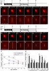
Figure 1.
Figure 1.
Confocal images of discrete TMR tracer injections and axonal projections after naris closure from 4 to 7 or 7 to 10 weeks. Time lines indicate weeks of naris closure and reopening, the time points when tracer injections were done (arrows), and the side of the nasal cavity that was blocked (the right side, R). A–L, TMR tracer injections (A–F) are shown in the dorsolateral OB of rI7→M71 mice with corresponding intrabulbar projection sites (G–L). Yellow lines represent the greatest lengths of injection and projection sites. A–L, Injection and projection series from juvenile mice following unilateral naris closure from 4 to 7 weeks of age (B, H), along with subsequent recovery periods of 3 weeks (C, I), 6 weeks (D, J), and 9 weeks (E, K) showing gradual refinement of the intrabulbar map. Control mice at 7 weeks (A, G) and 16 weeks (F, L) of age are included for comparison. M–X, Similar series in adult mice show tracer injections (M–R) and corresponding projection sites (S–X) of animals blocked from 7 to 10 weeks (N, T) and those followed by 3 weeks (O, U), 6 weeks (P, V) or 9 weeks (Q, W) of recovery as well as 10-week (M, S) and 19-week (R, X) controls, showing that the expansion and gradual re-refinement of intrabulbar projections does not have a critical period. Graphs illustrate mean projection to injection ratios (±SEM) and the trend of projection broadening and refinement observed in both age groups (Y) along with the significant differences observed after the 4–7 and 7–10 week blocks (*p < 0.001, t = 15.40 and 12.04, respectively) as compared to age matched controls (Z). Significant differences were also found after 4–7 or 7–10 weeks of naris closure with 3 weeks (*p < 0.001, t = 6.29 and 10.23, respectively) or 6 weeks (*p < 0.001, t = 4.34 and *p < 0.001, t = 3.90, respectively) but not 9 weeks (p = 0.44, t = 0.78 and p = 0.16, t = 1.44, respectively) of recovery (Z). R, Right side; L, left side; M, mitral cell layer. Scale bars, 50 μm.
Figure 2.
Figure 2.
Photomicrographs of the lateral turbinates of P2-IRES-taulacZ mice. A–L, Mice served as controls (A, B, E, F) or underwent reversible naris closure from 7 to 10 weeks (C, D, G, H), or from 7 to 10 weeks with 3 weeks (I, K) or 6 weeks (J, L) of recovery. Lateral turbinates in the experimental side (right) of the nasal cavity are shown in C, D, I, and J, while contralateral control (left) turbinates are shown in G, H, K, and L. M, Graph shows the average number of lacZ+ cells in turbinate II (±SEM), revealing a significant reduction on the experimental side in the blocked group (*p < 0.01; t = 4.5) but no significant difference in cell number after naris block and reopening for 3 or 6 weeks or in controls. These results suggest that removing the block leads to a relatively rapid upregulation of Lac-Z+ cells on the previously occluded side. L, Left; R, right. Scale bar (in L): A, C, E, G, 500 μm; B, D, F, H, I–L, 350 μm.
Figure 3.
Figure 3.
Confocal images of β-gal-ir lateral glomeruli in P2-IRES-taulacZ mice. B, F, Images of lateral P2 β-gal-ir glomeruli are shown (green) with a DAPI counterstain (blue) and illustrate an average 55.7% difference in glomerular volume found in mice that experienced reversible naris closure from 7 to 10 weeks of age (n = 4). A, C, D, E, G, H, This difference was not found in age-matched controls (n = 3) (A, E) or in mice that underwent naris closure from 7 to 10 weeks followed by 3 weeks (n = 4) (C, G) or 6 weeks (n = 3) (D, H) of normal olfactory stimulation. I, Graph shows average P2 glomerular volume measurements (±SEM) and reveals a significant reduction on the experimental (right) side following naris closure from 7 to 10 weeks (*p < 0.001; t = −6.0), but no left–right differences after the block and 3 or 6 weeks of recovery. L, Left; R, right; NL, nerve layer; GLM, glomerular layer; Glom., glomerular. Scale bar, 50 μm.
Figure 4.
Figure 4.
Three-dimensional reconstructions of intrabulbar axonal tufts. Flattened images of confocal stacks of intrabulbar axonal projection sites are shown with corresponding 3D reconstructions below in each case. A–D, Representative confocal images and reconstructions are shown from 10-week-old controls (A), mice that experienced naris closure from 7 to 10 weeks (B), and mice that underwent naris block followed by 3 weeks (C) or 6 weeks (D) of normal levels of afferent activity. Scale bar, 50 μm.
Figure 5.
Figure 5.
Activity-dependent changes in synaptoporin-ir puncta density within the EPL and IPL. A–H, Confocal images of synaptoporin immunostaining in OB sections from 10 week control mice (A, E), mice that underwent naris closure from 7 to 10 weeks (B, F), and mice that experienced naris closure followed by 3 weeks (C, G) or 6 weeks (D, H) of recovery. The white dotted line in A–D delineates the mitral cell layer. Boxed regions of the IPL of A–D are shown in higher magnification in E–H. I, Graph shows the quantification of synaptoporin-ir puncta densities in the EPL and IPL. Naris closure led to a decrease in the density of synaptoporin-ir puncta in the IPL (*p < 0.05; t = 4.51), and this decrease persisted in mice that underwent olfactory deprivation followed by 3 weeks of recovery (*p < 0.05; t = 3.51), but not in the group that recovered for 6 weeks. In addition, no significant differences were observed in synaptoporin-ir puncta densities within the EPL. Scale bar (in H): A–D, 50 μm; E–H, 30 μm.
Figure 6.
Figure 6.
Model of intrabulbar map plasticity. A, In the adult OB, isofunctional glomeruli (shown in green) are connected by STC axons (shown in red) that reciprocally project to the region of the IPL located below the “partner” glomerulus. B, If odorant-induced activity is blocked by naris closure, glomerular size becomes reduced and intrabulbar projections broaden. C, Reopening the naris for 3 weeks results in the recovery of glomerular volume; however, the intrabulbar projections remain broad. D, Reopening the naris and recovery for ∼6–9 weeks allows the intrabulbar map to return to control levels of precision.

References

    1. Alonso M, Viollet C, Gabellec MM, Meas-Yedid V, Olivo-Marin JC, Lledo PM. Olfactory discrimination learning increases the survival of adult-born neurons in the olfactory bulb. J Neurosci. 2006;26:10508–10513. - PMC - PubMed
    1. Baker H, Morel K, Stone DM, Maruniak JA. Adult naris closure profoundly reduces tyrosine hydroxylase expression in mouse olfactory bulb. Brain Res. 1993;614:109–116. - PubMed
    1. Belford GR, Killackey HP. The sensitive period in the development of the trigeminal system of the neonatal rat. J Comp Neurol. 1980;193:335–350. - PubMed
    1. Belluscio L, Lodovichi C, Feinstein P, Mombaerts P, Katz LC. Odorant receptors instruct functional circuitry in the mouse olfactory bulb. Nature. 2002;419:296–300. - PubMed
    1. Bozza T, Feinstein P, Zheng C, Mombaerts P. Odorant receptor expression defines functional units in the mouse olfactory system. J Neurosci. 2002;22:3033–3043. - PMC - PubMed

Publication types

MeSH terms

LinkOut - more resources