. 2010 Jul 8;466(7303):248-52.
doi: 10.1038/nature09151.

A random cell motility gradient downstream of FGF controls elongation of an amniote embryo

Affiliations

A random cell motility gradient downstream of FGF controls elongation of an amniote embryo

Bertrand Bénazéraf et al. Nature. .

Abstract

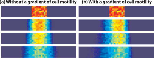
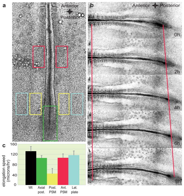
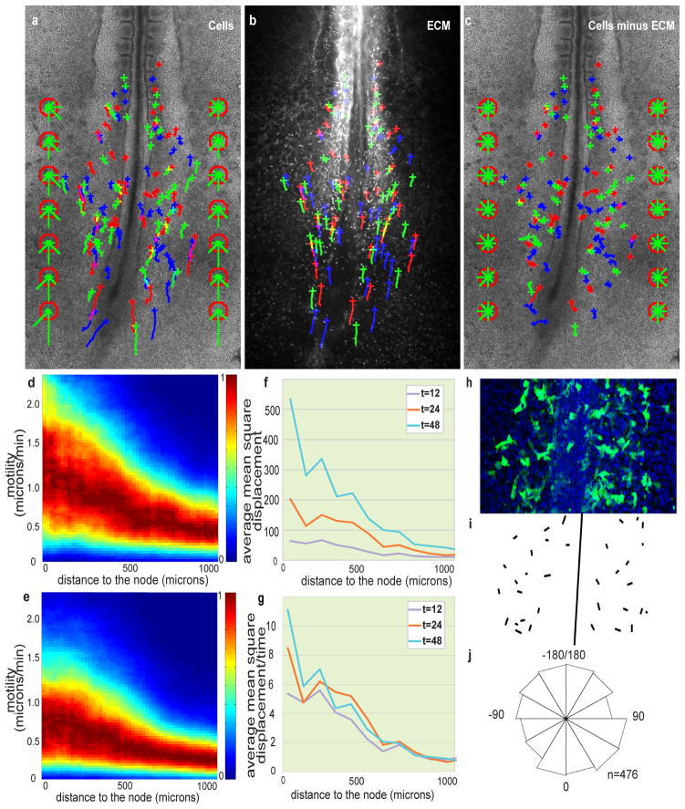
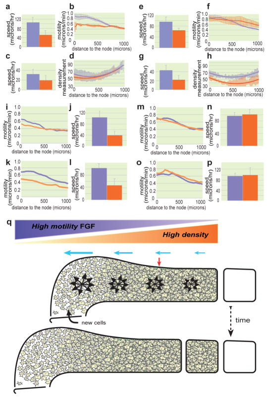
Vertebrate embryos are characterized by an elongated antero-posterior (AP) body axis, which forms by progressive cell deposition from a posterior growth zone in the embryo. Here, we used tissue ablation in the chicken embryo to demonstrate that the caudal presomitic mesoderm (PSM) has a key role in axis elongation. Using time-lapse microscopy, we analysed the movements of fluorescently labelled cells in the PSM during embryo elongation, which revealed a clear posterior-to-anterior gradient of cell motility and directionality in the PSM. We tracked the movement of the PSM extracellular matrix in parallel with the labelled cells and subtracted the extracellular matrix movement from the global motion of cells. After subtraction, cell motility remained graded but lacked directionality, indicating that the posterior cell movements associated with axis elongation in the PSM are not intrinsic but reflect tissue deformation. The gradient of cell motion along the PSM parallels the fibroblast growth factor (FGF)/mitogen-activated protein kinase (MAPK) gradient, which has been implicated in the control of cell motility in this tissue. Both FGF signalling gain- and loss-of-function experiments lead to disruption of the motility gradient and a slowing down of axis elongation. Furthermore, embryos treated with cell movement inhibitors (blebbistatin or RhoK inhibitor), but not cell cycle inhibitors, show a slower axis elongation rate. We propose that the gradient of random cell motility downstream of FGF signalling in the PSM controls posterior elongation in the amniote embryo. Our data indicate that tissue elongation is an emergent property that arises from the collective regulation of graded, random cell motion rather than by the regulation of directionality of individual cellular movements.


Figures

Box 1 Figure 1
Box 1 Figure 1
Simulation results at t=0,50,100,250
Figure 1
Figure 1. Role of caudal PSM in embryo elongation
a, Position of the ablations of the posterior PSM (yellow rectangles), lateral plate mesoderm (light blue rectangles), caudal axial tissue (green rectangle) and anterior PSM (red rectangles) in a stage HH11 embryo. b, Snapshot of different time points (during an 8-hour time period) showing elongation (red lines) of a wild type (Wt) embryo starting at stage HH11. The left red line denotes the level of somite 3, while the right red line denotes the level of Hensen’s node. c, Elongation speed measurements. Error bars indicate the standard deviations. The black histogram corresponds to the elongation rate of non-ablated Wt embryos.
Figure 2
Figure 2. Posterior-to-anterior decreasing gradient of random motility in the PSM
a–c, H2B-GFP electroporated embryos injected with labelled anti-Fibronectin antibodies. Crosses represent cell position at t = 0; trajectories are represented by green, blue and red lines. The green bars in the red octagons represent cell directionality (see Supplementary Methods). a, Tracking of cellular movements. b, Tracking of ECM movement (Fibronectin). c, Cell movements after subtraction of ECM motion. d, e, Cellular motility along the AP axis with respect to a somite; node position is on the left; normalized probabilities for different motilities are colour coded (see Supplementary Methods). d, Gradient of cellular motility. e, Gradient of cellular motility relative to the ECM. f, Analysis of the mean square displacement of cells relative to the ECM along the AP axis as a function of time (in min.). g, Mean square displacement normalized by time (in min.). h–j, Orientation of cellular protrusions relative to the AP axis. h, Ventral view of the caudal part of an embryo electroporated with membrane-GFP. i, Representation of the orientations of major lamelliform protrusion. j, Rose diagram representing the distribution of the angles between the major cellular lamelliform protrusion and the AP body axis (n = 476 cells from 14 different embryos, 0 degrees corresponds to a protrusion parallel to the AP axis and pointing toward the tail).
Figure 3
Figure 3. Effect of FGF signaling, cell movement and cell proliferation on axis elongation
a–p, Control (lavender) and treated (orange) conditions. a, e, j, l, n, p, Quantification of elongation. c, g, Convergence analysis. b, f, i, k, m, o, Mean cellular motility analysed along the AP axis. d, h, Analysis of PSM cellular density along the AP axis. The solid line represents a curve fitted to the mean value and the standard deviation is represented by the corresponding light-colored bars (n=4 or 5 for each condition). a–d, Inducible FGFR1dn electroporation. e–h, FGF8 electroporation. i,j, Y27632 treatment. k,l, Blebbistatin treatment. m,n, Aphidicolin treatment. o,p, Mitomycin treatment. b,f mean of two [for FGFR1dn] or three embryos [for FGF8]; errors bars represent the standard deviation). q, Model of the control of elongation by a gradient of random cellular motion in the PSM. Dorsal view of a schematic representation of the left PSM at two consecutive stages of embryo elongation. While new cells are entering the PSM, the gradient of random motility (lavender gradient; black arrow clusters) opposed to the gradient of cellular density (orange gradient) creates a directional bias in elongation (blue arrows) toward the posterior part of the tissue. This posterior expansion induces the convergence of the PSM tissue (red arrow).

References

    1. Dubrulle J, Pourquie O. fgf8 mRNA decay establishes a gradient that couples axial elongation to patterning in the vertebrate embryo. Nature. 2004;427:419–422. - PubMed
    1. Delfini MC, Dubrulle J, Malapert P, Chal J, Pourquie O. Control of the segmentation process by graded MAPK/ERK activation in the chick embryo. Proc Natl Acad Sci USA. 2005;102:11343–11348. - PMC - PubMed
    1. Keller R, Shook D, Skoglund P. The forces that shape embryos: physical aspects of convergent extension by cell intercalation. Physical biology. 2008;5:15007. - PubMed
    1. Lawson A, Schoenwolf GC. New insights into critical events of avian gastrulation. Anat Rec. 2001;262:238–252. - PubMed
    1. Voiculescu O, Bertocchini F, Wolpert L, Keller RE, Stern CD. The amniote primitive streak is defined by epithelial cell intercalation before gastrulation. Nature. 2007;449:1049–1052. - PubMed

Publication types

Substances