. 2010 Jun 29;8(6):e1000407.
doi: 10.1371/journal.pbio.1000407.

Human mucosal associated invariant T cells detect bacterially infected cells

Affiliations

Human mucosal associated invariant T cells detect bacterially infected cells

Marielle C Gold et al. PLoS Biol. .

Abstract

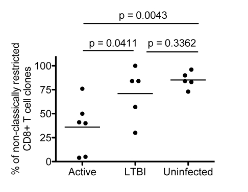
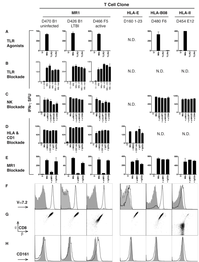
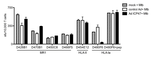
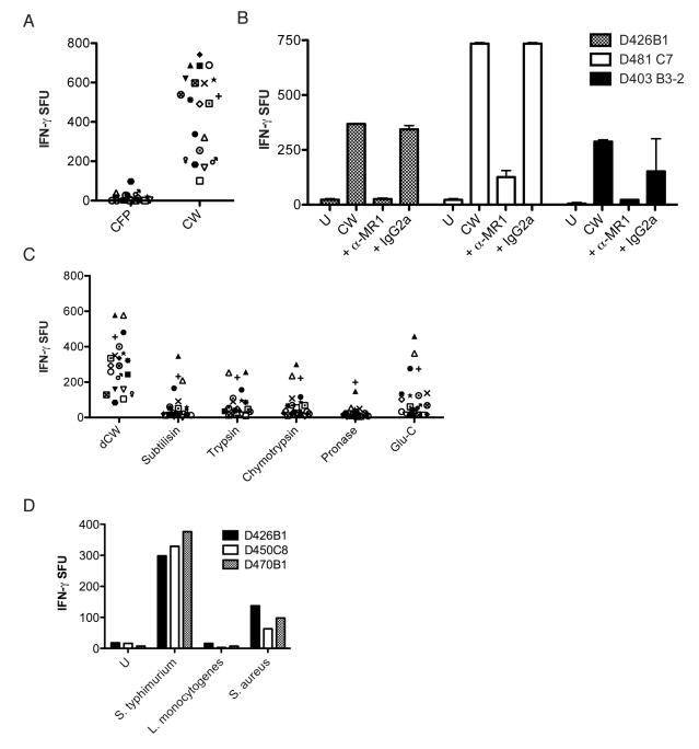
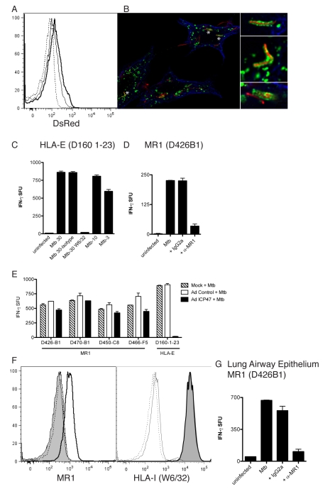
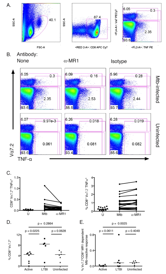
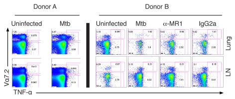
Control of infection with Mycobacterium tuberculosis (Mtb) requires Th1-type immunity, of which CD8+ T cells play a unique role. High frequency Mtb-reactive CD8+ T cells are present in both Mtb-infected and uninfected humans. We show by limiting dilution analysis that nonclassically restricted CD8+ T cells are universally present, but predominate in Mtb-uninfected individuals. Interestingly, these Mtb-reactive cells expressed the Valpha7.2 T-cell receptor (TCR), were restricted by the nonclassical MHC (HLA-Ib) molecule MR1, and were activated in a transporter associated with antigen processing and presentation (TAP) independent manner. These properties are all characteristics of mucosal associated invariant T cells (MAIT), an "innate" T-cell population of previously unknown function. These MAIT cells also detect cells infected with other bacteria. Direct ex vivo analysis demonstrates that Mtb-reactive MAIT cells are decreased in peripheral blood mononuclear cells (PBMCs) from individuals with active tuberculosis, are enriched in human lung, and respond to Mtb-infected MR1-expressing lung epithelial cells. Overall, these findings suggest a generalized role for MAIT cells in the detection of bacterially infected cells, and potentially in the control of bacterial infection.


Conflict of interest statement

The authors have declared that no competing interests exist.

Figures

Figure 1
Figure 1. LDA of Mtb-reactive CD8+ T-cell clones.
Scatter plot demonstrating the proportion of NC restricted CD8+ T-cell clones obtained from individuals in the active, LTBI, and uninfected groups. Each symbol represents the average frequency from all clones screened from an individual donor (Table 1), which was categorized as NC restricted. The nonparametric Mann Whitney one-tailed t-test was used to assess statistical significant differences between groups. Significant differences were detected between active and uninfected groups (p = 0.0043) between the active and LTBI groups (p = 0.0411), but not between the LTBI and uninfected groups (p = 0.3362).
Figure 2
Figure 2. Mtb-specific NC CD8+ T cells are restricted by MR1.
(A–E) Results of ELISPOT assays shown as IFN-γ spot forming units (SFU)/10,000 T cells in response to DCs (25,000/well) treated as described. (A) TLR agonist stimulation of DCs does not stimulate Mtb-reactive NC-restricted clones. DCs were treated (24 h) with TLR agonists specific for TLR2 (lipoteichoic acid, 10 µg/ml) and TLR4 (LPS; 100 ng/ml) at concentrations known to induce activation and cytokine production by DCs . (B) TLR2 (5 µg/ml) or TLR4 (10 µg/ml) blocking antibodies were added to DCs that were uninfected or infected 1 h prior to the addition of Mtb-reactive NC T-cell clones. (C) Mtb-infected DCs were incubated with blocking antibodies (5 µg/ml) to NKG2D, ULBP1, MICA, CD94 for 1 h prior to the addition of the T-cell clones. (D) The pan HLA–I (W632) and CD1a, b, c, and d blocking antibodies were added to Mtb-infected DCs prior to the addition of T cells. (E) DCs infected with Mtb overnight were incubated with anti-MR1 blocking antibody (clone 26.5) or a mouse IgG2a isotype control (both at 5 µg/ml) for 1 h prior to the addition of T cells. (F–H) Cell surface phenotypic analyses of MR1-restricted clones and control clones. For cell surface detection, cells were incubated with antibodies specific for Vα7.2 (clone 3C10) (F), or CD8α, CD8β (G), or CD161 (H), and analyzed by flow cytometry. For (F) and (H), filled histograms represent the isotype control, bold lines represent antibody-specific staining. Columns 1, 2, and 3 represent MR1-restricted clones from different TB exposure groups: D470B1 (uninfected), D426B1 (latent), D466F5 (active), respectively. Column 4 represents HLA-E restricted clone D160 1–23 . Column 5 represents HLA-B08-restricted clone D480C6 specific for the Mtb antigen CFP-103–11. Column 6 represents CD4+ HLA-II–restricted clone D454E12 specific for the Mtb antigen CFP-10. Error bars represent the mean and standard error from duplicate wells. N.D., not done.
Figure 3
Figure 3. MR1-restricted recognition of Mtb-infected cells is TAP-independent.
DCs autologous to D454 and expressing HLA-B08 were transduced with either a control adenoviral vector or adenoviral ICP47 using lipofectamine 2000. After 16 h, DCs were washed and either left uninfected, infected with Mtb, or pulsed with HLA-B08 specific peptide CFP103–11. Following overnight incubation, T cells were added (10,000) to DCs (25,000/well) and IFN-γ production was assessed by ELISPOT. Results are representative of three independent assays. No responses were detected from T cells incubated with uninfected DCs with or without adenoviral vectors. Error bars represent the mean and standard error from duplicate wells.
Figure 4
Figure 4. MR1 presents a protein-containing antigen from the mycobacterial cell wall.
(A) CFP or CW from the Mtb strain H37Rv were added (5 µg/ml) to DCs (25,000/well) for 1 h prior to the addition of one of 21 NC clones (5,000/well) followed by IFN-γ ELISPOT assay. (B) DCs (25,000) loaded with CW overnight were incubated with anti-MR1 blocking antibody (clone 26.5) or a mouse IgG2a isotype control (both at 5 µg/ml) for 1 h prior to the addition of T-cell clones (10,000/well). (C) dCW from Mtb was treated with proteases (subtilisin, trypsin, chymotrypsin, pronase, Glu-C) and added (5 µg/ml) to DCs (25,000/well) for 1 h before the addition of 21 NC clones (5,000/well) that were tested for their ability to produce IFN-γ in an ELISPOT assay. Reversed phase- high performance liquid chromatography (RP-HPLC) chromatogram analyses were used to confirm the inactivation of proteases. No responses were detected in the absence of DCs. (D) DCs were infected with S. typhimurium, L. monocytogenes, and S. aureus for 1 h with a calculated moi of 145, 6, and 15, respectively. DCs were washed, antibiotics added, and DCs (25,000) were incubated with three different Mtb-reactive MAIT-cell clones (10,000/well) that were tested for their ability to produce IFN-γ in an ELISPOT assay. Results shown are similar to a minimum of three independent experiments where S. typhimurium, L. monocytogenes, and S. aureus were tested at a variety of moi ranging from 5 to 150.
Figure 5
Figure 5. Characterization of MR1-dependent recognition of human lung epithelial cells.
(A) Flow cytometric analysis of A549 cells left uninfected (dashed line) or infected with Mtb H37Rv dsRED at an moi of 10 (solid line) or an moi of 30 (bold line). (B) A549 cells were infected with Mtb (dsRED-expressing H37Rv moi of 30∶1) and incubated overnight. Cells were washed, fixed (4% paraformaldehyde), and permeabilized (0.2% saponin) before being stained for expression of Lamp1 (green) and tubulin (blue). Images were acquired on a high-resolution wide-field Core DV system (Applied Precision) with a Nikon Coolsnap ES2 HQ. One 0.5-µm Z-section is shown. Asterisks indicate magnified regions. (C) A549 cells uninfected or infected with Mtb over a range and at the moi of 30 in the presence of anti-pan HLA-I blocking antibody (W6/32) or the IgG2a isotype control (2 µg/ml each) before being used as APCs for HLA-E–restricted T-cell clone D160 1–23 in the IFN-γ ELISPOT assay. (D) A549 cells uninfected or infected with Mtb (moi 30) were used as APCs for T-cell clone D426B1 in the presence of anti-MR1 blocking antibody or the IgG2a isotype control (2 µg/ml each) in the IFN-γ ELISPOT assay. Similar results were obtained from four different MR1-restricted T-cell clones. (E) A549 cells were infected with either control vector or adenoviral ICP47. After 16 h, A549 cells were washed and left uninfected or were infected with Mtb (moi 30). Following overnight incubation, T cells were added (10,000) and IFN-γ production was assessed by ELISPOT. Results are representative of three independent assays. Error bars represent the mean and standard error from duplicate wells. (F) Flow cytometric analysis of A549 cells that were left uninfected or infected with Mtb (moi of 30). The filled histogram represents cell surface MR1 (left panel) or HLA-I (right panel) staining on uninfected cells. All lines represent cell surface staining performed on Mtb-infected A549 cells (moi 30). Dashed line, Alexa 647 secondary control; solid line, msIgG2a isotype control; bold line, cell-surface staining of MR1 (26.5)(left panel) , or HLA-I (W6/32) (right panel). Cell surface expression of HLA-I is identical on uninfected and infected A549 cells. (G) Human primary large airway epithelial cells were infected with Mtb (moi 30) and used as APCs (25,000/well) for MR1-restricted T-cell clone D426B1 (15,000/well) in the IFN-γ ELISPOT assay in the presence of no, anti-MR1 (26.5) or IgG2a isotype control antibodies (2 µg/ml final each). Similar results were obtained in three independent experiments.
Figure 6
Figure 6. Ex vivo analysis of Mtb-reactive MAIT cells from human peripheral blood CD8+ T cells.
T cells were isolated from PBMC using the negative pan-T cell isolation kit and then depleted of CD4+ T cells. The resulting T cells were incubated overnight with A549 cells that were either Mtb-infected (moi 30) or left uninfected in the presence of no, anti-MR1, or IgG2a control antibody. Golgi Stop was added for the final 6 h of the assay. Cells were surface stained for expression of the Vα7.2 TCR then fixed and permeabilized before staining for TNF-α and CD8 expression. Controls performed to test for specific TNF-α staining showed no background responses (unpublished data). (A) Gating strategy: lymphocyte gate (left), CD8 gate (middle), Vα7.2 (y-axis), and TNF-α expression (x-axis) of the CD8 gate (right). The number on the upper left of the right panel represents the frequency of CD8+ T cells that expressed the Vα7.2 TCR. The number in the box on the right represents the frequency of CD8+ T cells that produced TNF-α in response to target cells. (B) Representative FACS analysis showing the conditions tested in the assay: Mtb-uninfected or infected A549 cells in the presence of anti-MR1 antibody (26.5) or IgG2a isotype control (2.5 µg/ml each). (C) Frequency of CD8+ cells that produced TNF-α in response to uninfected, Mtb-infected, or MR1-blocked Mtb-infected A549 cells that either coexpressed the Vα7.2 TCR (left) or not (right). (D) Frequency of CD8+ T cells that expressed the Vα7.2 TCR. Statistically significant differences were observed between the active and LTBI groups. (E) Frequency of MR1-blocked CD8+ T cells that coexpressed Vα7.2 and TNF-α in response to Mtb-infected cells. Statistically significant differences were observed between the active and uninfected groups. Horizontal lines in (D) and (E) represent medians. The nonparametric Mann-Whitney one-tailed t-test was used to assess statistical significant differences between groups.
Figure 7
Figure 7. Mtb-reactive MAIT cells in the human lung.
Single cell suspensions were prepared from the lung and adjacent LNs with minor modifications . The intracellular cytokine staining assay was performed using magnetic-bead purified CD8+ T cells from the lung and LNs as described in Figure 6 legend. Only in the case of donor B were anti-MR1 or IgG2a isotype control antibodies added (2.5 µg/ml).

References

    1. WHO. Geneva: World Health Organization; 2008. Global tuberculosis control - surveillance, planning, financing.
    1. Flynn J. L, Chan J. Immunology of tuberculosis. Annu Rev Immunol. 2001;19:93–129. - PubMed
    1. Lewinsohn D. A, Heinzel A. S, Gardner J. M, Zhu L, Alderson M. R, et al. Mycobacterium tuberculosis-specific CD8+ T cells preferentially recognize heavily infected cells. Am J Respir Crit Care Med. 2003;168:1346–1352. - PubMed
    1. Lewinsohn D. A, Winata E, Swarbrick G. M, Tanner K. E, Cook M. S, et al. Immunodominant tuberculosis CD8 antigens preferentially restricted by HLA-B. PLoS Pathog. 2007;3:1240–1249. doi: 10.1371/journal.ppat.0030127. - DOI - PMC - PubMed
    1. Lewinsohn D. M, Briden A. L, Reed S. G, Grabstein K. H, Alderson M. R. Mycobacterium tuberculosis-reactive CD8+ T lymphocytes: the relative contribution of classical versus nonclassical HLA restriction. J Immunol. 2000;165:925–930. - PubMed

Publication types

MeSH terms

Substances