. 2010 Jun 29;5(6):e11333.
doi: 10.1371/journal.pone.0011333.

Disruption of Dnmt1/PCNA/UHRF1 interactions promotes tumorigenesis from human and mice glial cells

Affiliations

Disruption of Dnmt1/PCNA/UHRF1 interactions promotes tumorigenesis from human and mice glial cells

Eric Hervouet et al. PLoS One. .

Abstract

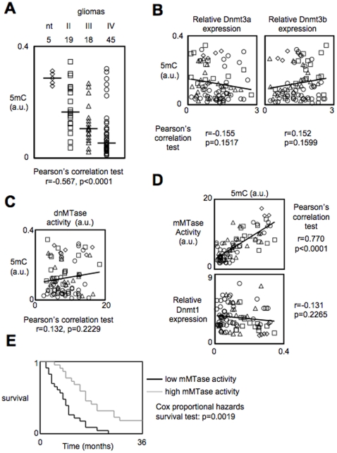
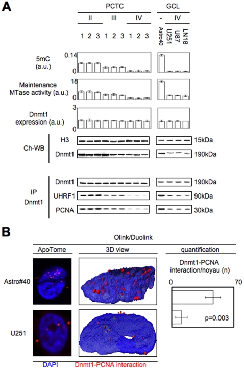
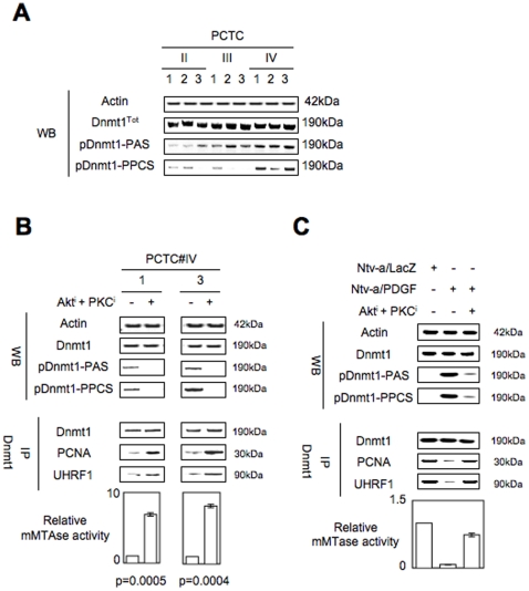
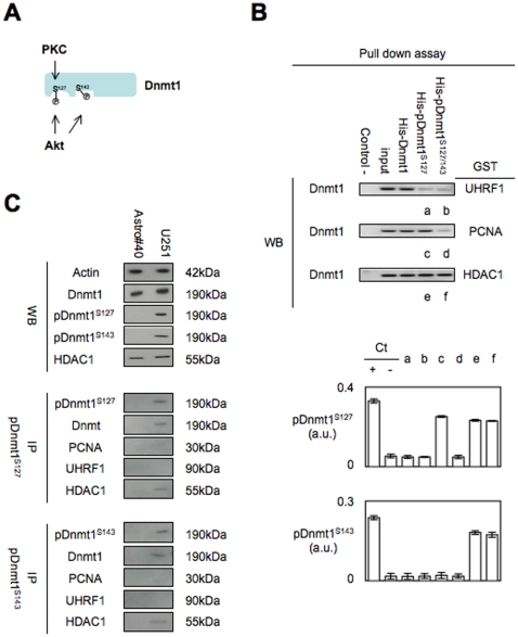
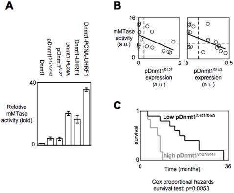
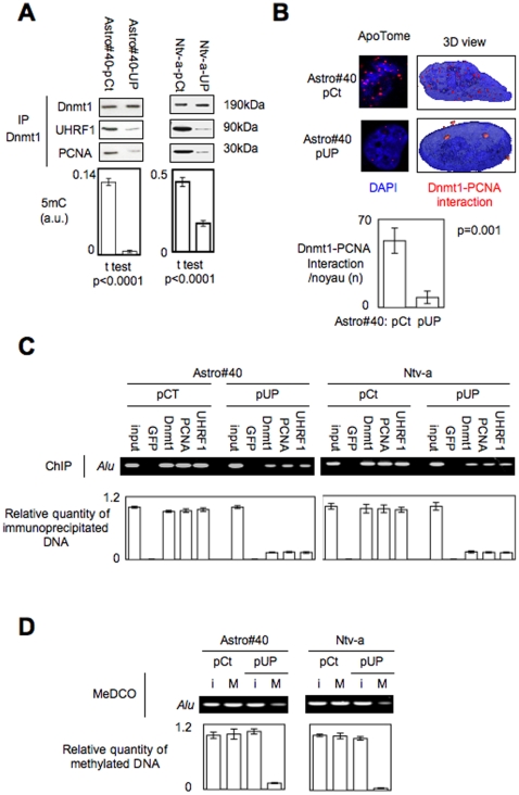
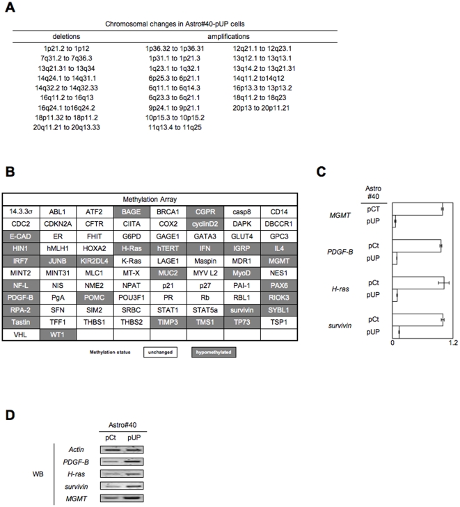
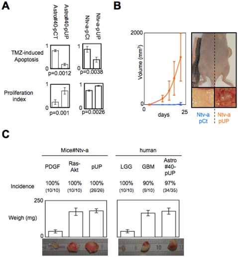
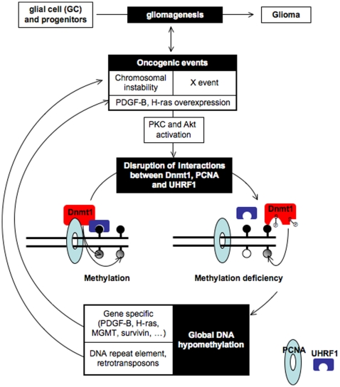
Global DNA hypomethylation is a hallmark of cancer cells, but its molecular mechanisms have not been elucidated. Here, we show that the disruption of Dnmt1/PCNA/UHRF1 interactions promotes a global DNA hypomethylation in human gliomas. We then demonstrate that the Dnmt1 phosphorylations by Akt and/or PKC abrogate the interactions of Dnmt1 with PCNA and UHRF1 in cellular and acellular studies including mass spectrometric analyses and the use of primary cultured patient-derived glioma. By using methylated DNA immunoprecipitation, methylation and CGH arrays, we show that global DNA hypomethylation is associated with genes hypomethylation, hypomethylation of DNA repeat element and chromosomal instability. Our results reveal that the disruption of Dnmt1/PCNA/UHRF1 interactions acts as an oncogenic event and that one of its signatures (i.e. the low level of mMTase activity) is a molecular biomarker associated with a poor prognosis in GBM patients. We identify the genetic and epigenetic alterations which collectively promote the acquisition of tumor/glioma traits by human astrocytes and glial progenitor cells as that promoting high proliferation and apoptosis evasion.


Conflict of interest statement

Competing Interests: The authors have declared that no competing interests exist.

Figures

Figure 1
Figure 1. The decrease of maintenance methyltransferase activity (mMTase) is correlated with the genome hypomethylation occurring during gliomagenesis, and confers poor prognosis in glioma patients.
(A) Correlation between the 5-methylcytosine number (5 mC) and tumor grade in a collection of 82 glioma and 5 non-tumor brain samples (nt). 5 mC was estimated by using the Methylamp Global DNA Methylation Quantification kit (Epigentek-Euromedex, France). Dotted lines represent the median of each parameter. (B) Correlation study between the 5-methylcytosine number (5 mC) and level expression of Dnmt3a (left) and Dnmt3b (right). (C) Correlation study between the 5-methylcytosine number (5 mC) and the de novo methyltransferase (dnMTase) calculated by using unmethylated DNA substrates in DMB assays as previously described . (D) Correlation study between the 5-methylcytosine number (5 mC) and the maintenance methyltransferase (mMTase) calculated by using hemi-methylated DNA substrates in DMB assays (Top). Correlation study between relative Dnmt1 expression and the 5-methylcytosine number (5 mC) (bottom). (E) Kaplan-Meier estimates time of survival between patients suffering from glioma presenting a high level of mMTase activity (grey line) and those whose tumors harbored a low level of mMTase activity (black line). In this figure, ⋄ represents 5 non-tumor brain samples, □ represents grade II astrocytomas/oligodendroglioma, ▵ represents grade III astrocytomas/oligodendroglioma, ○ represents grade IV astrocytomas/GBM.
Figure 2
Figure 2. Disruption of the Dnmt1/PCNA and Dnmt1/UHRF1 interactions in gliomagenesis.
(A) 5-methylcytosine (5 mC) number, maintenance methyltransferase (mMTase) activity, Dnmt1 expression, Dnmt1 recruitment on DNA and Dnmt1/PCNA/UHRF1 interaction in a panel of PCTC issued from the different grade of glioma. 5 mC was estimated by using the Methylamp Global DNA Methylation Quantification kit (Epigentek-Euromedex, France). mMTase activity and Dnmt1 expression was measured from nuclear extract obtained by using the EpiQuik Nuclear Extraction Kit I (Euromedex, France). Immunoprecipitations are performed by using the Catch and Release® v2.0 Reversible Immunoprecipitation System (Millipore, France). (B) Use of proximity ligation in situ assay (P-LISA) to monitor the disruption of the Dnmt1-PCNA interactions. Nucleus/DNA are in blue and Dnmt1-PCNA interaction in red. Quantification was performed from the analysis of 100 cells of three independent experiments.
Figure 3
Figure 3. The disruption of the Dnmt1/PCNA/UHRF1 interactions is associated with the phsphorylation of Dnmt1 by Akt and/or PKC.
(A) Visualization, in primary cultured tumor cells (PCTC) of different grade of glioma, of the phosphorylation level of Dnmt1 by using antibodies recognizing the phospho-(Ser/Thr) Akt substrate (pDnmt1-PAS, Ozyme, Cell Signal#9614, France) and the the phospho-(Ser) PKC substrate (pDnmt1-PPCS, Ozyme, Cell Signal#2261, France). (B) Effect of the Akt and PKC inhibition on the phosphorylation level of Dnmt1, the Dnmt1-PCNA-UHRF1 interactions and on the mMTAse activity. (Akti: 0.1 µM Calbiochem#124005, France; PKCi or Go6893, 0.5 µM Calbiochem#124005, France). (C) Effect of the constitutive activation of Akt and PKC in Ntv-a/PDGF cells on the phosphorylation level of Dnmt1, the Dnmt1-PCNA-UHRF1 interactions and on the mMTAse activity.
Figure 4
Figure 4. The phsophorylation of the Dnmt1 at S127 and/orS143 decrease the capacity of the protein to interact with PCNA and UHRF1.
(A) Schematic representation of the Akt and PKC-induced phosphorylation of Dnmt1 according to the results obtained by mass spectrometric approach. (B) Effect of the phosphorylation of the recombinant Dnmt1 (Methylation Ltd, Port Orange, Florida) by Akt and PKC on the Dnmt1-PCNA, Dnmt1-UHRF1 and Dnmt1-HDAC1 interactions. Pull-down assays were performed by using the GST/His Tag Protein Interaction Pull-Down Kits (Thermo Scientic, France). Fusion protein purification gel is illustrated in Supplemental data S5. The phoshorylation level of the Dnmt1 interacting with GST-PCNA, GST-UHRF1 and GST-HDAC1 was analyzed by ELISA method. Ct+: Dnmt1R phosphorylated by Akt and/or PKC, Ct-: Dnmt1R. (C) Western blot analyses were realized to monitor the expression level of Dnmt1, pDnmt1S127, pDnmt1S143 and HDAC1, Actin was used as control. IP experiments were realized by using the Catch and Release® v2.0 Reversible Immunoprecipitation System (Millipore, France) with 4 µg of antibody. Western blot of IP experiments illustrate the results obtained by adding the immunoprecipitate obtained from 3 immunoprecipitations performed with 500 µg of proteins.
Figure 5
Figure 5. pDnmt1S127/S143 and/or pDnmt1S127 catalyze low mMTase activity in comparison with Dnmt1/PCNA/UHRF1 and is hallmark associated with poor prognosis in glioma.
(A) Analysis of the mMTAse activity catalyzed by the Dnmt1, the Akt-mediated phosphorylation of the Dnmt1 (pDnmt1-PAS), the PKC-mediated phosphorylation of the Dnmt1 (pDnmt1-PPCS), in presence of equimolar quantity of PCNA, (Dnmt1-PCNA), or UHRF1 (Dnmt1-UHRF1) or PCNA and UHRF1 (Dnmt1-PCNA-UHRF1). mMTAse activities were assessed by DMB assay according to Yokochi and Robertson (2004). (B) Correlation study between the mMTase activity and the expression level of pDnmt1S127 and pDnmt1S143 harbored by 16 GBM. ○ represents grade IV astrocytomas/GBM. (C) Kaplan-Meier estimates time of survival between patients suffering from glioma presenting a high expression level of pDnmt1S127 and pDnmt1S143 (grey line) and those whose tumors harbored a low expression level of pDnmt1S127 and pDnmt1S143 (black line).
Figure 6
Figure 6. The disruption of the Dnmt1/PCNA/UHRF1 interactions promotes the global DNA hypomethylation in astrocytes (Astro#40) and in glial precusor cells (Ntv-a).
(A) Monitoring by immunoprecipitation of Dnmt1 and ELISA of the effect of the expression of the UP protein” (a chimera protein composed by the 163–171 amino-acids of PCNA and the 526–614 amino-acids of UHRF1) on the Dnmt1/PCNA/UHRF1 interactions and on the level of 5-methylcytosine (5 mC). (B) Use of proximity ligation in situ assay (P-LISA) to monitor the “UP”-induced disruption of the Dnmt1-PCNA interactions. Nucleus/DNA are in blue and Dnmt1-PCNA interaction in red. Quantification was performed from the analysis of 100 cells of three independent experiments. (C) Impact of the “UP”-induced disruption of the Dnmt1-PCNA-UHRF1 interactions on the co-recruitment of Dnmt1, PCNA and UHRF1 on Alu, a DNA repeat element. Chromatin Immunoprecipitation (ChIP) was performed by using the EZ-ChIP (Millipore, France). For each point, the relative quantity of immunoprecipitated DNA is obtained by using input as reference. (D) Impact of the “UP”-induced disruption of the Dnmt1-PCNA-UHRF1 interactions on the methylation status of Alu by coupling the Methylated DNA COllection and PCR amplification (MeDCO) via the use of the MethylCollector Ultra kit (Active Motif, France). (I:input: M:Methylated and collected DNA).
Figure 7
Figure 7. The disruption of the Dnmt1/PCNA/UHRF1 interactions, in astrocytes (Astro#40) and in glial precusor cells (Ntv-a), promotes chromosomal instability and gene-specifc hypomethylation.
(A) Description of chromosomal changes in Astro#40-UP cells via the realization of CGH array. (B) Description of changes in the methylation profil Astro#40-UP cells via the realization of Methylation Array (Ozyme-Panomics, France). (C) Validation by Methylated DNA Immunoprecipitation (MeDIP) and PCR amplification (MeDIP) of the hypomethylation of the MGMT, PDGF-B, H-ras and survivin genes in Astro40#UP cells. (I:input: M:Methylated and collected DNA). For each point, the relative quantity of immunoprecipitated DNA is obtained by using input as reference. (D) Expression level of the MGMT, PDGF-B, H-ras and survivin proteins in Astro#40 and Astro40#UP cells via western blot analysis.
Figure 8
Figure 8. Impact of the “UP”-induced disruption of the Dnmt1/PCNA/UHRF1 interactions on hallmarks of cancer in Astro#40 and Ntv-a cells.
(A) Comparison of the temozolomide-induced apopto-sensitivity (via the measure of DEVDase activity) and the proliferation index between the Astro#40 and Astro#40-UP cells and between the Ntv-a and Ntv-a/UP cells. DEVDase activity was assessed as previously described. Proliferation index was evaluated by quantify the cells number. (B) Tumorigenicity test of the Ntv-a and Ntv-a/UP cells. (C) Comparison of the tumorogenicity of the Ntv-a/UP cells with the one of the Ntv-a/PDGF and Ntv-a/Ras-Akt cells (left) and of the tumorogenicity of the Astro#40-UP cells with the one of the primary cultured tumor cells (PCTC) obtained from low-grade glioma (LGG) or glioblastoma multiforme (GBM) (right). Pictures are representative of tumors obtained after cells injection.
Figure 9
Figure 9. Schematic representation of gliomagenesis induced by the disruption of the Dnmt1-PCNA-UHRF1 interactions.

References

    1. Gama-Sosa M, Slagel V, Trewyn R, Oxenhandler R, Kuo K, et al. The 5-methylcytosine content of DNA from human tumors. Nucleic Acids Res. 1983;11:6883–6894. - PMC - PubMed
    1. Feinberg A, Vogelstein B. Hypomethylation distinguishes genes of some human cancers from their normal counterparts. Nature. 1983;301:89–92. - PubMed
    1. Esteller M. Epigenetics in cancer. N Engl J Med. 2008;358:1148–1159. - PubMed
    1. Hoffmann M, Schulz W. Causes and consequences of DNA hypomethylation in human cancer. Biochem Cell Biol. 2005;83:296–321. - PubMed
    1. Ehrlich M, Woods C, Yu M, Dubeau L, Yang F, et al. Quantitative analysis of associations between DNA hypermethylation, hypomethylation, and DNMT RNA levels in ovarian tumors. Oncogene. 2006;25:2636–2645. - PMC - PubMed

Publication types