

Frequency of developmental dental anomalies in the Indian population
- PMID: 20613914
- PMCID: PMC2897859
Frequency of developmental dental anomalies in the Indian population
Abstract
Objectives: To evaluate the frequency of developmental dental anomalies in the Indian population.
Methods: This prospective study was conducted over a period of 1 year and comprised both clinical and radiographic examinations in oral medicine and radiology outpatient department. Adult patients were screened for the presence of dental anomalies with appropriate radiographs. A comprehensive clinical examination was performed to detect hyperdontia, talon cusp, fused teeth, gemination, concrescence, hypodontia, dens invaginatus, dens evaginatus, macro- and microdontia and taurodontism. Patients with syndromes were not included in the study.
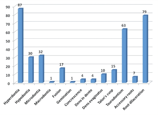
Results: Of the 20,182 patients screened, 350 had dental anomalies. Of these, 57.43% of anomalies occurred in male patients and 42.57% occurred in females. Hyperdontia, root dilaceration, peg-shaped laterals (microdontia), and hypodontia were more frequent compared to other dental anomalies of size and shape.
Conclusions: Dental anomalies are clinically evident abnormalities. They may be the cause of various dental problems. Careful observation and appropriate investigations are required to diagnose the condition and institute treatment.
Keywords: Dental anomalies; Hyperdontia; Microdontia; Taurodontism.
Figures
References
-
- Neville DW, Damm DD, Allen CM, Bouquot JE. Oral and Maxillofacial Pathology. 2nd ed. Philadelphia, PA: Elsevier; 2005. Abnormalities of teeth; pp. 49–89.
-
- Neville DW, Damm DD, Allen CM, Bouquot JE. Color Atlas of Clinical Oral Pathology. 2nd ed. Baltimore, MD: Williams and Wilkins; 1991. pp. 62–64.
-
- Vasudev SK, Goel BR. Endodontic management of dens evaginatus of maxillary central incisors: A rare case report. J Endod. 2005;31:67–70. - PubMed
-
- Juan JS, Jiménez-Rubio A. Talon cusp affecting permanent maxillary lateral incisors in 2 family members. Oral Surg Oral Med Oral Pathol Oral Radiol Endod. 1999;88:90–92. - PubMed
-
- Romito LM. Concrescence: report of a rare case. Oral Surg Oral Med Oral Pathol Oral Radiol Endod. 2004;97:325–327. - PubMed
LinkOut - more resources
Full Text Sources