Effect of different polymerization methods on the cytotoxicity of dental composites
- PMID: 20613917
- PMCID: PMC2897862
Effect of different polymerization methods on the cytotoxicity of dental composites
Abstract
Objectives: The aim of this study was to compare the cytotoxic effects of various dental composites polymerized with two different curing units.
Methods: Disc-shaped test samples of composites Filtek Z250, Filtek A110, Filtek P60, Filtek Supreme, and SDI Rok were polymerized using one quartz tungsten halogen (QTH) and one light emitting diode (LED) light curing unit (LCU), namely Optilux 501 (QTH) and Elipar Freelight 2 (LED). L-929 mouse fibroblast cultures (3x10(4) cells/ml) were incubated with the samples in 96 well culture plates for evaluation after 8, 24, 48, 72 h. At the end of each period, the cells were counted and examined under a light microscope, and a 3-[4,5-dimethylthiazol-2-yl]-2,5-diphenyltetrazolium bromide (MTT) assay was performed. The degree of cytotoxicity for each sample was determined according to the reference value represented by the cells in a control group (a culture without sample).
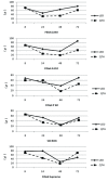
Results: A significant 3 factor interaction occurred among LCUs, composites, and time factors (P<.005). In general, the test materials cured with the LED LCU demonstrated higher cell survival rates when compared with those cured with halogen LCUs.
Conclusions: This study shows that polymerization of dental composites with a light emitting diode LCU positively influences the L-929 mouse fibroblast cell viability.
Keywords: Cytotoxicity; Dental composite; Light curing units.
Figures
References
-
- Davidson CL, Feilzer AJ. Polymerization shrinkage and polymerization shrinkage stress in polymer-based restoratives. J Dent. 1997;25:435–440. - PubMed
-
- Moon HJ, Lee YK, Lim BS, Kim CW. Effects of various light curing methods on the leachability of uncured substances and hardness of a composite resin. J Oral Rehabil. 2004;31:258–264. - PubMed
-
- Engelmann J, Leyhausen G, Leibfritz D, Geurtsen W. Metabolic effects of dental resin components in vitro detected by NMR spectroscopy. J Dent Res. 2001;80:869–875. - PubMed
-
- Geurtsen W. Substances released from dental resin composites and glass ionomer cements. Eur J Oral Sci. 1998;106:687–695. - PubMed
-
- Ferracane JL, Greener EH. Fourier transform infrared analysis of degree of polymerization in unfilled resins--methods comparison. J Dent Res. 1984;63:1093–1095. - PubMed
LinkOut - more resources
Full Text Sources


