. 2010 Jun 29;5(6):e11379.
doi: 10.1371/journal.pone.0011379.

Methylation of the tumor suppressor protein, BRCA1, influences its transcriptional cofactor function

Affiliations

Methylation of the tumor suppressor protein, BRCA1, influences its transcriptional cofactor function

Irene Guendel et al. PLoS One. .

Abstract

Background: Approximately half of hereditary breast cancers have mutations in either BRCA1 or BRCA2. BRCA1 is a multifaceted tumor suppressor protein that has implications in processes such as cell cycle, transcription, DNA damage response and chromatin remodeling. This multifunctional nature of BRCA1 is achieved by exerting its many effects through modulation of transcription. Many cellular events are dictated by covalent modification of proteins, an important mechanism in regulating protein and genome function; of which protein methylation is an important posttranslational modification with activating or repressive effects.

Methods/principal findings: Here we demonstrate for the first time that BRCA1 is methylated both in breast cancer cell lines and breast cancer tumor samples at arginine and lysine residues through immunoprecipitation and western blot analysis. Arginine methylation by PRMT1 was observed in vitro and the region of BRCA1 504-802 shown to be highly methylated. PRMT1 was detected in complex with BRCA1 504-802 through in vitro binding assays and co-immunoprecipitated with BRCA1. Inhibition of methylation resulted in decreased BRCA1 methylation and alteration of BRCA1 binding to promoters in vivo as shown through chromatin immunoprecipitation assays. Knockdown of PRMT1 also resulted in increased BRCA1 binding to particular promoters in vivo. Finally, following methylation inhibition, Sp1 was found to preferentially associate with hypo-methylated BRCA1 and STAT1 was found to preferentially associate with hyper-methylated BRCA1.

Conclusions/significance: These results suggest that methylation may influence either the ability of BRCA1 to bind to specific promoters or protein-protein interactions which alters the recruitment of BRCA1 to these promoters. Thus, given the importance of BRCA1 to genomic stability, methylation of BRCA1 may ultimately affect the tumor suppressor ability of BRCA1.


Conflict of interest statement

Competing Interests: The authors have declared that no competing interests exist.

Figures

Figure 1
Figure 1. BRCA1 is methylated at both arginine and lysine residues in breast cancer cell lines.
(a) Predicted BRCA1 methylation sites generated by Memo: Methylation Modification Prediction Server (http://www.bioinfo.tsinghua.edu.cn/~tigerchen/memo.html) R = Arginine and K = Lysine. (b) Two milligram of whole cell protein extracts from MCF-7 and MDA-MB-231 cells were immunoprecipitated with either BRCA1(C-20) or rabbit normal IgG antibody, separated on a 4–20% gel by SDS-PAGE, and western blotted with antibodies against K-methyl, R-methyl and BRCA1(C-20). Input represents 1/10 of immunoprecipitated material. Results are representative of three independent experiments. (c) One milligram of whole cell protein extracts from MDA-MB-231 and BRCA1-null UWB1.289 cells were immunoprecipitated with either BRCA1(C-20) or rabbit normal IgG antibody, separated on a 4–20% gel by SDS-PAGE, and western blotted with antibodies against R-methyl and BRCA1(C-20) in order to validate observed methylated bands. Densitometry of R-methylation was normalized to amount of detected immunoprecpipitated BRCA1, background subtracted based on IgG counts. (d) One milligram of whole cell protein extracts from synchronized MDA-MB-231 cells were immunoprecipitated with either BRCA1(C-20) or rabbit normal IgG antibody, separated on a 4–20% gel by SDS-PAGE, and western blotted with antibodies against R-methyl and BRCA1(C-20). Densitometry of R-methylation was normalized to amount of detected immunoprecpipitated BRCA1, background subtracted based on IgG counts. Input represents 1/10 of immunoprecipitated material. (e) Whole cell protein extracts from synchronized MDA-MB-231 cells were separated on a 4–20% gel by SDS-PAGE, and western blotted with antibodies against Cyclin B(H-433), Cyclin D1(M-20), Cyclin E(C-19), cdk4(H-303) and actin(C-11).
Figure 2
Figure 2. Methylation of BRCA1 in ex vivo breast tumor samples.
(a) Solid breast tumor tissue was ground with a mortar and pestle in the presence of liquid nitrogen to create a powdered tissue. RIPA buffer was added to the powdered tissue, the sample vortexed for 60 seconds, and placed on ice for 45 minutes. Samples were homogenized with a syringe and needle, followed by centrifugation at 14,000 g for 10 minutes. Two milligram of whole cell protein extracts from four different breast tumor samples (BT1-4) were immunoprecipitated with either BRCA1(C-20) or rabbit normal IgG antibody, separated on a 4–20% gel by SDS-PAGE, and western blotted with an anti-K methyl, anti-R methyl and anti-BRCA1 antibodies. Results are representative of two independent experiments. Characteristics of tumor tissues are as follows: BT1: infiltrating ductal carcinoma, age 55, African American, ER negative, PR negative, HER2 negative, p53 negative. BT2: infiltrating lobular carcinoma, age 83, Caucasian, ER positive, PR negative, HER2 negative, p53 negative. BT3: variant papillary serous type of ductal carcinoma, age 54, African American, ER negative, PR negative, HER2 negative, p53 negative. BT4: infiltrating ductal carcinoma, age 68, Caucasian, ER negative, PR negative, HER2 negative, p53 positive. (b) Matched breast tumor tissue was processed as described. Four milligram of whole cell protein extract were immunoprecipitated with either BRCA1(C-20) or rabbit normal IgG antibody, separated on a 4–20% gel by SDS-PAGE, and western blotted with an anti-R methyl and anti-BRCA1 antibodies. Input represents 1/5 of immunoprecipitated material.
Figure 3
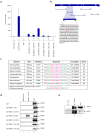
Figure 3. PRMT1 methylates and associates with BRCA1.
(a) GST-BRCA1 constructs (0.5 µg) and core histones (1 µg) were incubated with purified recombinant PRMT1 enzyme (0.2 µg) in the presence of 0.55 µCi SAdenosyl- L-[methyl-3H] methionine. GST was used as negative control and all GST-BRCA1 construct methylation levels normalized to background GST methylation levels. An average result of two independent experiments run in triplicates is shown. (b) The amino acid sequence of BRCA1 504–802 with arginine (R) residues within the 540–696 minimal region highlighted in blue and bolded, and the predicted methylated arginine residue highlighted in red, underlined and bolded. (c) Multiple sequence alignment generated by Clustal W of four non-human primates and two rodent BRCA1 full length sequences. Score represents pairwise alignment against full length human BRCA1. Amino acid residues are color-coded according to physical properties: basic (magenta), small hydrophobic (red) and hydroxyl + amine + basic (green). Consensus symbols for degree of conservation observed is represented by “*” (residues in column are identical in all seven sequences), “:” (conserved substitutions observed), and “.” (semi-conserved substitutions observed). (d) Two milligram of HeLa whole cell protein extract was incubated with 0.5 µg GST-BRCA1 constructs, beads were washed twice with TNE150 + 0.1% NP-40 and once with TNE50 + 0.1% NP-40, separated on a 4–20% gel by SDS-PAGE, and probed with an antibody against PRMT1. Input represents 1/10 of immunoprecipitated material. (e) Two milligram of HeLa whole cell protein extract were immunoprecipitated with anti-BRCA1 and anti-IgG antibodies, beads were washed twice with TNE150 + 0.1% NP-40 and once with TNE50 + 0.1% NP-40, separated on a 4–20% by SDS-PAGE, and probed with an anti-PRMT1 antibody. Input represents 1/10 of immunoprecipitated material. Results are representative of two independent experiments.
Figure 4
Figure 4. Methylation status of BRCA1 alters BRCA1-DNA interactions at specific promoters in vivo.
(a) MDA-MB-231 cells were treated with AdOx (30 µM) in order to observe BRCA1 methylation inhibition upon treatment. Two milligram of MDA-MB-231 whole cell protein extract was immunoprecipitated with anti-BRCA1 and anti-IgG antibodies, separated on a 4–20% gel by SDS-PAGE, and probed with anti-K methyl antibody. Blot was stripped and reprobed with anti-BRCA1 antibody. Input represents 1/10 of immunoprecipitated material. Results are representative of two independent experiments. (b) MDA-MB-231 cells were treated with AdOx (30 µM) for 48 hours prior to being collected for ChIP analysis. Antibodies used for ChIP were anti-BRCA1 (10 µg), anti-IgG (10 µg), and anti-histone H3-phosphorylated at S10 (H3-pS10, 5 µg). PCR products were run on a 2% agarose gel and visualized with ethidium bromide staining. Results are representative of two independent experiments.
Figure 5
Figure 5. Decreased levels of PRMT1 alters BRCA1 promoter binding in vivo.
(a) HeLa cells were transfected with different concentrations of PRMT1 siRNA (10, 25, 50 nM) following manufacturer's instructions. Results are representative of two independent experiments. (b) HeLa cells transfected with 50 nM Luc or PRMT1 siRNA were collected for ChIP analysis. Anti-BRCA1 (10 µg), anti-IgG (10 µg), and anti-histone H3-phosphorylated at S10 (H3-pS10, 5 µg) antibodies were used for ChIP analysis. PCR products were run on a 2% agarose gel and visualized with ethidium bromide staining. Results are representative of two independent experiments.
Figure 6
Figure 6. BRCA1 methylation status alters protein-protein interactions at the 504-802 region.
(a) Schematic of BRCA1 504-802 primary sequence depicting important protein-protein interactions and domains that could be affected by the methylation of this region. (b) MDA-MB-231 cells were treated with AdOx (30 µM) in order to observe BRCA1 methylation inhibition upon treatment. Two milligram of MDA-MB-231 whole cell protein extract was immunoprecipitated with anti-BRCA1 or anti-IgG antibodies, separated on a 4-20% gel by SDS-PAGE, and western blotted using antibodies against Sp1 and BRCA1 proteins. Input represents 1/10 of immunoprecipitated material. Results are representative of two independent experiments. (c) MDA-MB-231 cells were treated with AdOx (30 µM) and whole cell extract separated on a 4-20% gel by SDS-PAGE, and probed with anti-Sp1 antibody. Densitometry was averaged from three independent immunoblots.

References

    1. Monteiro AN, August A, Hanafusa H. Evidence for a transcriptional activation function of BRCA1 C-terminal region. Proc Natl Acad Sci U S A. 1996;93:13595–13599. - PMC - PubMed
    1. Glover JN, Williams RS, Lee MS. Interactions between BRCT repeats and phosphoproteins: tangled up in two. Trends Biochem Sci. 2004;29:579–585. - PubMed
    1. Scully R, Anderson SF, Chao DM, Wei W, Ye L, et al. BRCA1 is a component of the RNA polymerase II holoenzyme. Proc Natl Acad Sci U S A. 1997;94:5605–5610. - PMC - PubMed
    1. Krum SA, Miranda GA, Lin C, Lane TF. BRCA1 associates with processive RNA polymerase II. J Biol Chem. 2003;278:52012–52020. - PubMed
    1. Anderson SF, Schlegel BP, Nakajima T, Wolpin ES, Parvin JD. BRCA1 protein is linked to the RNA polymerase II holoenzyme complex via RNA helicase A. Nat Genet. 1998;19:254–256. - PubMed

Publication types