

Comparative study of the expression of p53, Ki67, E-cadherin and MMP-1 in verrucous hyperplasia and verrucous carcinoma of the oral cavity
- PMID: 20614262
- PMCID: PMC2807515
- DOI: 10.1007/s12105-007-0029-y
Comparative study of the expression of p53, Ki67, E-cadherin and MMP-1 in verrucous hyperplasia and verrucous carcinoma of the oral cavity
Abstract
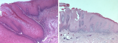
Context: Oral verrucous carcinoma (OVC) and oral verrucous hyperplasia (OVH) may be clinically and histologically similar. Problems separating these lesions are compounded by poorly oriented tissue sections and biopsies failing to demonstrate lesional margins.
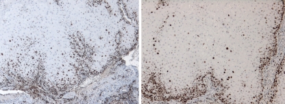
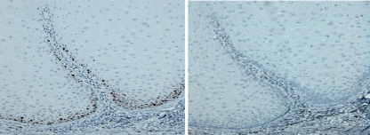
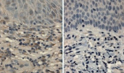
Objective: To distinguish OVC from OVH utilizing an immunohistochemical panel (p53, matrix metalloproteinase-1, E-cadherin, Ki67) shown to be useful in differentiating pseudoepitheliomatous hyperplasia from oral squamous cell carcinoma of the head and neck.
Materials: Twenty-eight cases of OVH and thirty-two cases of OVC studied. Diagnoses were confirmed by two pathologists. Formalin-fixed, paraffin-embedded archival material was used for immunohistochemistry (avidin-biotin immunoperoxidase technique).
Results: More diffuse nuclear staining of p53 and Ki67 was detected in the OVC cases compared to the OVH cases (P < 0.001). There was statistically significant increased staining within adjacent stromal cells for matrix metalloproteinase-1 (P < .05) in the OVC cases. E-cadherin demonstrated diffuse membranous staining in both groups.
Conclusion: Ki67, p53, and MMP-1 demonstrated significant staining trends. Although a properly oriented hematoxylin-eosin-stained section including normal marginal tissue is considered to be the gold standard for differentiation of OVH and OVC, this immunohistochemistry panel may serve as a useful diagnostic adjunct in difficult cases.
Keywords: Immunohistochemistry; Verrucous carcinoma; Verrucous hyperplasia.
Figures
References
-
- Zarovnaya E, Black C. Distinguishing pseudoepitheliomatous hyperplasia from squamous cell carcinoma in mucosal biopsy specimens from the head and neck. Arch Pathol Lab Med. 2005;129(8):1032–6. - PubMed
-
- Yantiss RK, Rosenberg MW, Odze RD. Utility of MMP-1, p53, E-cadherin, and collagen IV immunohistochemical stains in the differential diagnosis of adenomas with misplaced epithelium versus adenomas with invasive adenocarcinoma. Am J Surg Pathol. 2002;26:206–15. doi: 10.1097/00000478-200202000-00007. - DOI - PubMed
-
- Johnson N, Franceschi S, Slootweg PJ. World Health Organization classification of tumours. Lyon: International Agency for Research on Cancer Press; 2005. Squamous cell carcinoma. Pathology and genetics: head and neck tumors; pp. 175–6.
Publication types
MeSH terms
Substances
LinkOut - more resources
Full Text Sources
Medical
Research Materials
Miscellaneous
