

doi: 10.1007/s12105-007-0020-7.
Epub 2007 Dec 1.
An update on Epstein-Barr virus and nasopharyngeal carcinogenesis
Affiliations
- PMID: 20614265
- PMCID: PMC2807523
- DOI: 10.1007/s12105-007-0020-7
Item in Clipboard
An update on Epstein-Barr virus and nasopharyngeal carcinogenesis
Head Neck Pathol.
2007 Dec.
No abstract available
Figures
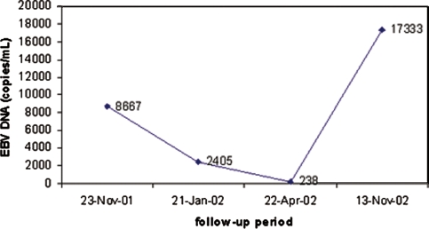
Patient with T3N1M0 nasopharyngeal carcinoma with 8,667 EBV-DNA copies/ml before combined chemoradiation. The EBV-DNA levels dropped to 238 copies/ml after completion of therapy but increased to 17,333 copies/ml after the development of pulmonary metastases. (Courtesy of Dr. Suzanne Kamel-Reid)

Nonkeratinizing nasopharyngeal carcinoma in-situ with strong EBERs expression by ISH (inset)
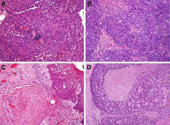
Surgical pathology of nasopharyngeal carcinoma. Nonkeratinizing carcinoma, differentiated-type composed of intermediate size cells with well defined eosinophilic cytoplasm and oval nuclei lacking prominent nucleoli. This case shows formation of a whorl (A). Nonkeratinizing carcinoma, undifferentiated-type is composed of large cells with amphophilic cytoplasm showing vesicular chromatin and prominent nucleoli (B). Keratinizing squamous carcinoma accompanied by carcinoma in-situ (C). Basaloid squamous cell carcinoma showing composed of small cells with peripheral palisading and formation of eosinophilic cylinders of basal lamina material (D)
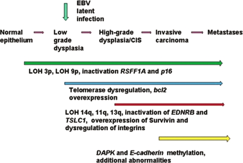
Proposed model of nasopharyngeal carcinogenesis with progressive accumulation of genetic and epigenetic abnormalities and corresponding histopathologic and clinical features
Similar articles
-
Quantitation of DNA methylation in Epstein-Barr virus-associated nasopharyngeal carcinoma by bisulfite amplicon sequencing.BMC Cancer. 2017 Jul 17;17(1):489. doi: 10.1186/s12885-017-3482-3. BMC Cancer. 2017. PMID: 28716111 Free PMC article.
-
Frequent hypermethylation of RASSF1A and TSLC1, and high viral load of Epstein-Barr Virus DNA in nasopharyngeal carcinoma and matched tumor-adjacent tissues.Neoplasia. 2005 Sep;7(9):809-15. doi: 10.1593/neo.05217. Neoplasia. 2005. PMID: 16229803 Free PMC article.
-
Nasopharyngeal carcinoma in two young brothers and its relationship with Epstein-Barr virus.Am J Otolaryngol. 1998 Sep-Oct;19(5):335-8. doi: 10.1016/s0196-0709(98)90009-6. Am J Otolaryngol. 1998. PMID: 9758184 No abstract available.
-
Epstein-Barr virus.IARC Monogr Eval Carcinog Risks Hum. 1997;70:47-373. IARC Monogr Eval Carcinog Risks Hum. 1997. PMID: 9612712 Free PMC article. Review. No abstract available.
-
Focus on nasopharyngeal carcinoma.Cancer Cell. 2004 May;5(5):423-8. doi: 10.1016/s1535-6108(04)00119-9. Cancer Cell. 2004. PMID: 15144950 Review. No abstract available.
Cited by
-
Characterization of the Variability of Epstein-Barr Virus Genes in Nasopharyngeal Biopsies: Potential Predictors for Carcinoma Progression.PLoS One. 2016 Apr 12;11(4):e0153498. doi: 10.1371/journal.pone.0153498. eCollection 2016. PLoS One. 2016. PMID: 27071030 Free PMC article.
-
Herpesviruses in Head and Neck Cancers.Viruses. 2020 Feb 3;12(2):172. doi: 10.3390/v12020172. Viruses. 2020. PMID: 32028641 Free PMC article. Review.
-
Role of infectious agents in the carcinogenesis of brain and head and neck cancers.Infect Agent Cancer. 2013 Feb 2;8(1):7. doi: 10.1186/1750-9378-8-7. Infect Agent Cancer. 2013. PMID: 23374258 Free PMC article.
-
A Systematic Review of Epstein-Barr Virus Latent Membrane Protein 1 (LMP1) Gene Variants in Nasopharyngeal Carcinoma.Pathogens. 2021 Aug 20;10(8):1057. doi: 10.3390/pathogens10081057. Pathogens. 2021. PMID: 34451521 Free PMC article. Review.
-
Implication of human herpesviruses in oncogenesis through immune evasion and supression.Infect Agent Cancer. 2014 Jan 20;9(1):3. doi: 10.1186/1750-9378-9-3. Infect Agent Cancer. 2014. PMID: 24438207 Free PMC article.
References
Publication types
MeSH terms
Substances
LinkOut - more resources
Full Text Sources
