. 2007 Sep;1(1):35-7.
doi: 10.1007/s12105-007-0018-1. Epub 2007 Nov 27.

Head and neck pathology-radiology classics: vagal paraganglioma

Affiliations

Head and neck pathology-radiology classics: vagal paraganglioma

Edina Paal et al. Head Neck Pathol. 2007 Sep.
No abstract available


Figures

Fig. 1
Fig. 1
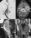
(A) Angiography. Lateral view (patient’s face is on left side of image) of the arterial phase of a digital subtraction angiogram with left common carotid artery injection shows early, intense enhancement of a vascular mass (arrow) that is causing anterior deviation of the carotid artery (arrowhead). (B) Intravenous contrast-enhanced axial CT. Large enhancing mass in the left carotid space, deep to the sternocleidomastoid muscle (tailed arrow), causing lateral deviation and compression of the slit-like left jugular vein compared to normal right jugular vein (arrowheads). In addition, the mass deviates the internal and external carotid arteries (arrows) anteriorly on the left compared to the normal position seen on the right. (C) Sagittal reformat of intravenous contrast-enhanced CT. Anterior deviation of the carotid arteries (arrow) by the large mass containing many irregular, nontapering tumor vessels (arrowheads). (D) MR, T1-weighted coronal image of the neck. Intermediate signal mass in the left neck displacing the flow void of the left common carotid artery which would normally be in the same plane as that on the right (tailed arrows). Irregular flow voids in high-flow tumor vessels are also seen within the mass (arrowheads)
Fig. 2
Fig. 2
Histology, H&E, 20×. Note the organoid growth pattern with zellballen and the rich vasculature in the delicate septa between them
Fig. 3
Fig. 3
(A) Chromogranin immunostain, 40×. Chief cells forming the zellballen react with Chromogranin A, while the surrounding sustentacular cells are negative (left). (B) S100 immunostain, 40×. As a mirror image, sustentacular cells react with S100 highlighting the contour of the zellballen (right)

References

    1. Hunt JL. Diseases of the paraganglia system. In: Thompson LDR, editors. Head and Neck Pathology (A volume in the fundation in diagnostic pathology series ed. Goldblum JR) Elsevier, Philadelhia, PA 2006: 551–62.
    1. Barnes L, Tse LLY, Hunt JL. Carotid body paraganglioma. In: Barnes EL, Evenson JW, Reichardt P, Sidransky D, editors. Pathology and genetics of head and neck tumours. Lyon, France: IARC Press; 2005. pp. 364–365.
    1. Rao AB, Koeller KK, Adair CF. From the archives of the AFIP: Paragangliomas of the head and neck: radiologic-pathologic correlation. Radiographics. 1999;19:1605–32. - PubMed
    1. Olsen WL, Dillon WP, Kelly WM, Norman D, Brant-Zawadzki M, Newton TH. MR imaging of paragangliomas. AJR Am J Roentgenol. 1987;148(1):201–4. - PubMed
    1. Boedecker CC, Ridder GJ, Schipper J. Paragangliomas of the head and neck: diagnosis and treatment. Fam Cancer. 2005;4:55–9. doi: 10.1007/s10689-004-2154-z. - DOI - PubMed

MeSH terms

LinkOut - more resources