

Case Reports
doi: 10.1007/s12105-008-0088-8.
Epub 2008 Oct 22.
Parathyroid adenoma
Affiliations
- PMID: 20614300
- PMCID: PMC2807581
- DOI: 10.1007/s12105-008-0088-8
Item in Clipboard
Case Reports
Parathyroid adenoma
Head Neck Pathol.
2008 Dec.
No abstract available
Figures
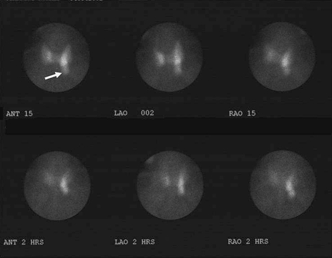
Fifty-seven year old man with mild hypercalcemia. 99mTc sestamibi scan demonstrates normal uptake by the thyroid gland with a focal area of pronounced uptake on the left (arrow). Over 2 h the thyroid uptake decreased to better reveal the prominent focal area of increased activity in the region of the lower left lobe of the thyroid consistent with parathyroid adenoma
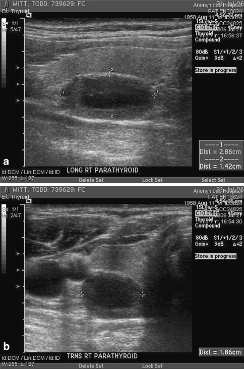
Sonographic image in the longitudinal (a) and transverse (b) plane demonstrates a hypoechoic parathyroid adenoma measuring 2.86 × 1.86 × 1.42 cm. Located centrally within the adenoma is an area of decreased echogenicity consistent with cystic degeneration
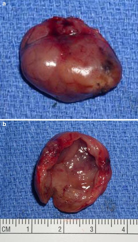
Gross specimen of a cystic parathyroid adenoma. (a) Exterior view shows a smooth surface with minimal hemorrhage and no significant adhesions. The specimen “shelled out” easily. (b) The mass is opened to demonstrate a smooth cyst wall lining
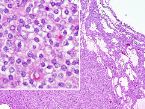
The low-power portion of this H&E stained section shows the hypercellular parathyroid adenoma with a cystic lumen indicated by the ** corresponding to the smooth cyst wall in Fig. 3b. High power examination (inset) depicts the proliferative parathyroid tissue to be composed of a population of chief cells. This parathyroid proliferation lacks the normal fat component
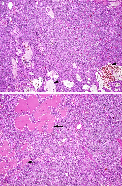
(a) H&E stained section shows a solid growth pattern interspersed with areas of cystic change (arrows). The cystic spaces are filled with a watery “proteinaceous” fluid as well as red blood cells. (b) A bright, eosinophilic material fills the “follicle-like” spaces (arrows) with peripheral scalloping reminiscent of the colloid-filled follicles seen in thyroid tissue
References
-
- Clark PB, Perrier ND, Morton KA. Detection of an intrathymic parathyroid adenoma using single-photon emission CT 99mTc sestamibi scintigraphy and CT. AJR Am J Roentgenol. 2005;184(Suppl 3):S16–8. - PubMed
-
- Thompson LDR. Benign neoplasms of the parathyroid gland. In: Thompson LDR, editor. Endocrine pathology, foundations in diagnostic pathology. Philadelphia: Churchill Livingstone/Elsevier; 2006. pp. 157–164.
Publication types
MeSH terms
Substances
LinkOut - more resources
Full Text Sources
