Case Reports
. 2008 Dec;2(4):328-32.
doi: 10.1007/s12105-008-0078-x. Epub 2008 Aug 22.

Kimura disease of the epiglottis

Affiliations
Case Reports

Kimura disease of the epiglottis

AbdullGaffar Badr et al. Head Neck Pathol. 2008 Dec.

Abstract

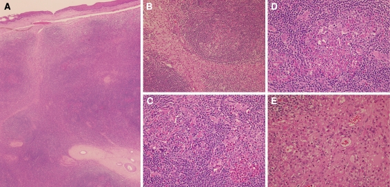
Kimura disease is a distinct clinicopathological entity of a benign chronic inflammatory disorder of unknown etiology. It is endemic in Oriental Asians, but sporadic and relatively rare in the West, both in whites and blacks alike. It usually presents as a mass lesion, most commonly in the head and neck region. It had for a long time been confused as synonymous with angiolymphoid hyperplasia with esinophilia. It can impose a challenging diagnosis both clinically and pathologically, especially in non-endemic areas with unusual sites involvement. Even though it is a benign lesion, it can be life-threatening in the epiglottis with a risk of airways obstruction. So far, one case had been reported in the epiglottis with upper respiratory tract obstruction. We report a similar case with a brief review of the literature.

Keywords: Epiglottis; Kimura disease.


Figures

Fig. 1
Fig. 1
(a) The epiglottis tissue is totally replaced by lymphoid proliferation, composed of lymphoid follicles with inter-follicular hyalinized blood vessels and fibrosed septae. Note the intact overlying surface squamous epithelium. (Hematoxylin & Eosin, original magnification × 40); (b) Reactive lymphoid follicle with maintained mantle zone and adjacent expanded inter-follicular septa with fibrosis and edema and with large number of esinophils. Note the absence of tingible body macrophages in the germinal centers. (Hematoxylin & Eosin, original magnification × 200); (c) Two lymphoid follicles are seen, one with aggregates of esinophils forming esinophilic microabscess and esinophilic folliculolysis and the other lymphoid follicle showing pink proteinaceous material with dismantling of the germinal center. (Hematoxylin & Eosin, original magnification × 400); (d) Lymphoid follicle showing prominent pink material deposition, esinophilic infiltration, proliferating small blood vessels and partial folliculolysis of the germinal center. Note the scattered multinucleated giant cells involving the germinal center and the interfollicular lymphoid tissue (H&E, original magnification × 400); (e) Inter-follicular septa with proliferating post-capillary venules with flat to plump endothelial cells, fibrosis and infiltrating esinophils. Note the absence of epithelioid-like endothelial cells (H&E, original magnification × 400)

References

    1. Abuel-Haija M, Hurford MT. Kimura disease. Arch Pathol Lab Med. 2007;131:650–1. - PubMed
    1. Chen H, Thompson LD, Aguilera NS, et al. Kimura’s disease: a clinicopathological study of 21 cases. Am J Surg Pathol. 2004;28:505–13. doi: 10.1097/00000478-200404000-00010. - DOI - PubMed
    1. Kuo TT, Shih LY, Chan HL. Kimura’s disease: Involvement of regional lymph nodes and distinction from angiolymphoid hyperplasia with esinophilia. Am J Surg Pathol. 1988;12:843–54. - PubMed
    1. Taskin M, Goksalan T, Kakani R, et al. Pathologic quiz case1. Pathologic diagnosis; Kimura’s disease. Arch Otolaryngol Head Neck Surg. 1996;122:893–4. - PubMed
    1. Urabe A, Tsuneyoshi M, Enjoji M. Epithelioid hemangioma vs. Kimura’s disease. A comparative clinicopathological study. Am J Surg Pathol. 1987;11:758–66. doi: 10.1097/00000478-198710000-00003. - DOI - PubMed

Publication types

MeSH terms