. 2010 Sep;48(9):554-8.
doi: 10.1002/dvg.20651.

Generation of mice with a conditional null allele for Wntless

Affiliations

Generation of mice with a conditional null allele for Wntless

April C Carpenter et al. Genesis. 2010 Sep.

Abstract

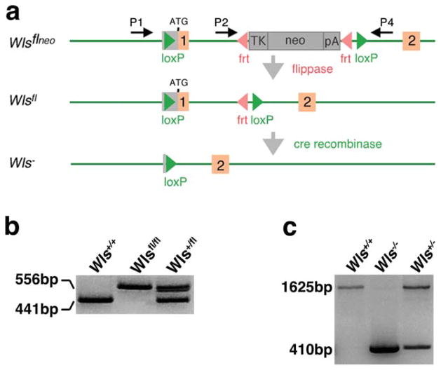
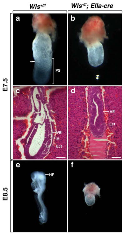
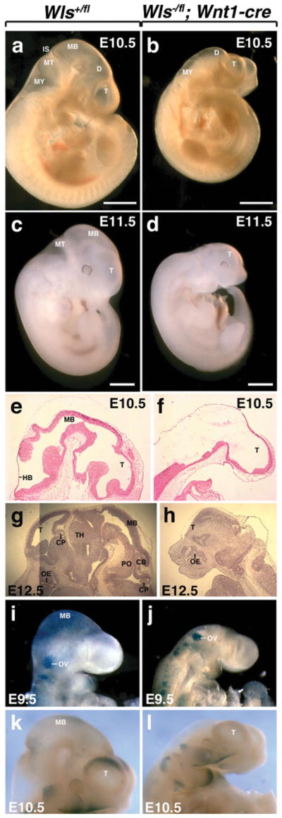
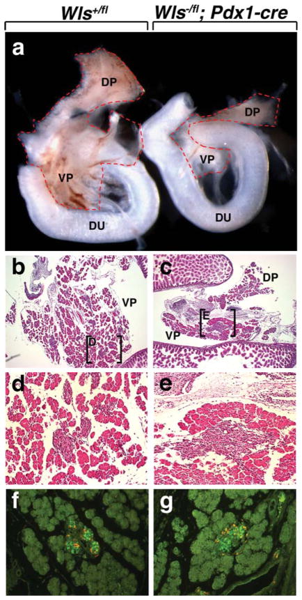
The Wnt-signaling pathway is necessary in a variety of developmental processes and has been implicated in numerous pathologies. Wntless (Wls) binds to Wnt proteins and facilitates Wnt sorting and secretion. Conventional deletion of Wls results in early fetal lethality due to defects in body axis establishment. To gain insight into the function of Wls in later stages of development, we have generated a conditional null allele. Homozygous germline deletion of Wls confirmed prenatal lethality and failure of embryonic axis formation. Deletion of Wls using Wnt1-cre phenocopied Wnt1 null abnormalities in the midbrain and hindbrain. In addition, conditional deletion of Wls in pancreatic precursor cells resulted in pancreatic hypoplasia similar to that previously observed after conditional β-catenin deletion. This Wls conditional null allele will be valuable in detecting novel Wnt functions in development and disease.


Figures

FIG. 1
FIG. 1
Generation of the Wls conditional allele. (a) Orange boxes correspond to exons. Red arrowheads are Frt sites. Green arrowheads denote LoxP sites. Recombination of the Frt sites flanking the Neo results in the floxed Wls allele. Recombination of the LoxP sites removes the ATG start site in exon 1 resulting in the Wls null (Wls) allele. P1, P2, and P4 represent the location of the primers used for Wls genotyping. (b) Successful Flp recombination was confirmed using PCR analysis to detect a 441-bp WT band and a 556 bp Wlsfl Neo deleted band (using primers P2 and P4). (c) Cre recombination was validated using PCR analysis to detect a 1,625-bp WT band and a 410-bp null band (using primers P1 and P4).
FIG. 2
FIG. 2
Deletion of germline Wls inhibits mouse embryogenesis. (a, b) Morphological analysis of embryos dissected at E7.5. The wild-type embryo has the characteristics of an early neural plate stage embryo with evidence of primitive streak formation. Wls−/− mutants resemble egg cylinders. The arrow denotes the embryonic-extra-embryonic junction at the anterior side. (c, d) Histological analysis of embryos dissected at E7.5. Wild-type embryos exhibit endoderm, mesoderm, and ectoderm. Wls−/− mutants display endoderm and ectoderm, but no mesoderm. Scale bars represent 100 μm. (e) Control embryo at E8.5 with a normal embryonic axis. (f) At E8.5 Wls−/− mutants still resemble egg cylinders. VE, visceral endoderm; M, mesoderm; Ect, ectoderm, PS, primitive streak; HF, head-fold.
FIG. 3
FIG. 3
Deletion of Wls in the dorsal midline of the diencephalon using Wnt1-cre. (a, b) Morphological analysis of embryos dissected at E10.5. Wls+/fl embryos exhibit a forebrain, midbrain and hindbrain. Wls/fl; Wnt1-cre embryos fail to develop a midbrain. (c) Wls+/fl embryos dissected at E11.5. (d) Wls/fl; Wnt1-cre embryos at E11.5 show defective development of midbrain, hindbrain and forebrain. Histologic comparison of WT (e, g) and mutant embryos (f, h). Mutant embryos display loss of midbrain and hindbrain formation at E10.5 (f). At E11.5, mutant embryos additionally lack a forebrain choroid plexus and display a modified forebrain (h). β-catenin/Wnt activity in the brain of Wls+/fl (i and k) and Wls/fl; Wnt1-cre (j and l) embryos stained for X-gal. Scale bars represent 1 mm. T, telencephalon; D, diencephalon; MB, midbrain; MT, metencephalon; MY, myencephalon; IS, isthmus; HB, hindbrain; CP, choroid plexus; OE, olfactory epithelium; TH, thalamus; PO, pons; CB, cerebellum; OV, otic vessicle.
FIG. 4
FIG. 4
Deletion of Wls in pancreatic precursor cells using Pdx1-cre. (a, b) Morphological analysis of pancreata dissected at E15.5. Both Wls+/fl and Wls/fl; Pdx1-cre pancreata exhibit a dorsal and ventral pancreas. Wls/fl; Pdx1 pancreata are severely hypoplastic (b). Histologic comparison of WT (b, d, f) and mutant pancreata (c, e, g). Low-power magnification of both Wls+/fl and Wls/fl; Pdx1-cre pancreata reveal the presence of acinar cells and Islets of Langerhans (b and c). Higher power magnification of the boxed regions in b and c (d and e, respectively). (f, g) E15.5 islets immunostained for insulin (green) and glucagon (orange). DP, dorsal pancreas; VP, ventral pancreas; Du, duodenum.

References

    1. Banziger C, Soldini D, Schutt C, Zipperlen P, Hausmann G, Basler K. Wntless, a conserved membrane protein dedicated to the secretion of Wnt proteins from signaling cells. Cell. 2006;125:509–522. - PubMed
    1. Bartscherer K, Pelte N, Ingelfinger D, Boutros M. Secretion of Wnt ligands requires Evi, a conserved transmembrane protein. Cell. 2006;125:523–533. - PubMed
    1. Belenkaya TY, Wu Y, Tang X, Zhou B, Cheng L, Sharma YV, Yan D, Selva EM, Lin X. The retromer complex influences Wnt secretion by recycling Wntless from endosomes to the trans-Golgi network. Dev Cell. 2008;14:120–131. - PubMed
    1. Brault V, Moore R, Kutsch S, Ishibashi M, Rowitch DH, McMahon AP, Sommer L, Boussadia O, Kemler R. Inactivation of the β-catenin gene by Wnt1-Cre-mediated deletion results in dramatic brain malformation and failure of craniofacial development. Development. 2001;128:1253–1264. - PubMed
    1. Danielian PS, Muccino D, Rowitch DH, Michael SK, McMahon AP. Modification of gene activity in mouse embryos in utero by a tamoxifen-inducible form of Cre recombinase. Curr Biol. 1998;8:1323–1326. - PubMed

Publication types