Review
. 2011 Mar 1;334(1-2):3-13.
doi: 10.1016/j.mce.2010.06.018. Epub 2010 Jul 6.

What are nuclear receptor ligands?

Affiliations
Review

What are nuclear receptor ligands?

Frances M Sladek. Mol Cell Endocrinol. .

Abstract

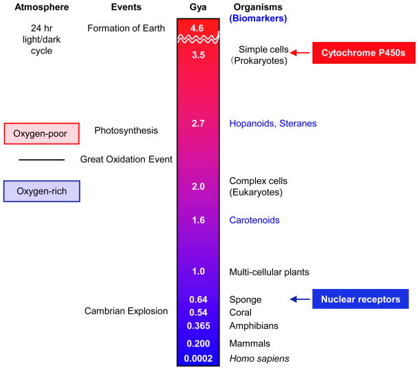
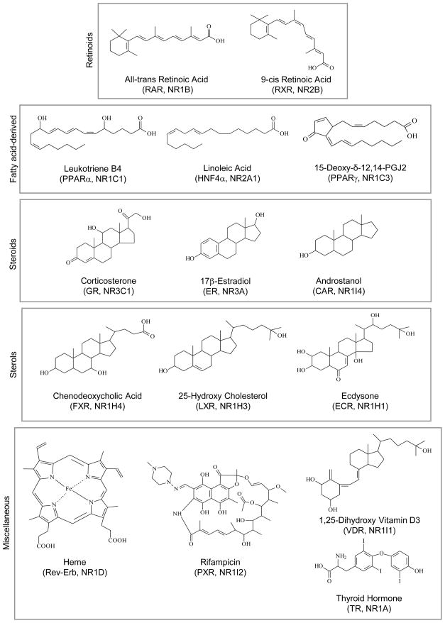
Nuclear receptors (NRs) are a family of highly conserved transcription factors that regulate transcription in response to small lipophilic compounds. They play a role in every aspect of development, physiology and disease in humans. They are also ubiquitous in and unique to the animal kingdom suggesting that they may have played an important role in their evolution. In contrast to the classical endocrine receptors that originally defined the family, recent studies suggest that the first NRs might have been sensors of their environment, binding ligands that were external to the host organism. The purpose of this review is to provide a broad perspective on NR ligands and address the issue of exactly what constitutes a NR ligand from historical, biological and evolutionary perspectives. This discussion will lay the foundation for subsequent reviews in this issue as well as pose new questions for future investigation.


Figures

Figure 1
Figure 1. Structure and mode of action of nuclear receptors
A. Domain structure of nuclear receptors (A–F). The DNA binding domain (DBD) consists of two zinc-binding motifs (Zn++) and often includes the hinge region. Shown is the crystallographic structure of the estrogen receptor ligand binding domain (LBD) bound to diethylstilbestrol (DES), a synthetic nonsteroidal estrogen that functions as a pure agonist, and the antagonist 4-hydroxy tamoxifen (4OHT) (dark blue), reprinted from Shaiu et al. (Shiau et al., 1998) with permission from Elsevier. AF1, AF2, activation function 1 and 2 (AF2, AF-2 helix); both contact co-regulatory molecules but AF-1 is typically ligand-independent while the AF-2 is ligand-dependent (colored magenta as helix 12). NRs have variable AF1 and hinge regions and F domains and range in size from ~450 to ~933 amino acids, although the majority are ~50 kDa. Many NR genes have multiple isoforms generated by alternative promoter usage and splice variants; some NR genes lack a DBD (not shown) (for a review of NR structures, see (Renaud and Moras, 2000)). B. Simplified diagram of classical NR action. In the absence of ligand, NRs recruit multi-subunit co-repressor complexes, which contain histone deacetylase activity (HDAC), to promoter regions of genes. Transcription of target genes is activated when ligand (hexagon) binds the NR and induces a conformation change in the LBD that recruits co-activator complexes that contain histone acetylase transferase activity (HAT). Not shown are other co-regulatory complexes needed for transcription such as chromatin remodeling and mediator complexes, and the general transcription machinery (see (Rosenfeld et al., 2006)).
Fig. 2
Fig. 2. Timeline of life on Earth
Shown is a highly simplified depiction of some of the major biological events and first appearance of various life forms, as evidenced in the fossil record, during the history of the Earth in billions of years (Gya) before present day. NRs are found in living relatives of the simplest animals (sponges, in the phylum Porifera), the first evidence of which appears ~635 Myr ago in the form of molecular fossils of the biomarker 24-isopropylcholestanes (Love et al., 2009). Cytochrome P450s, mono-oxygenases that bind heme, are in every living organism, including prokaryotes (Nelson, 2009). Biomarkers for bacteria (hopanoids, 2700 Myr ago), eukaryotes (steranes such as stigmastane and cholestane, 2700 Myr ago) and purple sulfur bacteria (carotenoids such okenone, 1600 Myr) are indicated in blue (structures are shown in Fig. 5); molecular fossils of steranes are often used as evidence that eukaryotes might have evolved much earlier than 2000 Myr ago (Brocks and Banfield, 2009; Brocks et al., 1999). The Earth’s atmosphere and oceans changed from an oxygen-poor to an oxygen-rich environment during the Great Oxygenation Event (~2300 Myr ago), ~300 million years after the advent of oxygenic photosynthesis by cyano- and other bacteria (~2600 Myr ago) (Reinhard et al., 2009). Cambrian explosion (520–550 Myr ago), the rapid appearance of most major groups of complex animals. Not indicated is the whole genome duplication of teleosteans (bony fishes) that occurred at 0.250 Gya and facilitated the expansion of the NR gene family.
Fig. 3
Fig. 3. Evolution of nuclear receptors: Relationship with morphological complexity of organisms?
Shown are the number of NR genes and their proposed role in various organisms representing different taxa, along with key features that distinguish each group from the one above it (complexity increases from top to bottom). Dashed line, there is some controversy over which organism – Trichoplax or Porifera (sponge) -- is at the base of the Metazoan Tree (Schierwater et al., 2009; Sperling et al., 2007). (*) Many nematode genes are expansions of a single NR gene (HNF4-like, NR2A). (**) Fish (zebrafish and fugu) NR numbers are higher than anticipated due to a recent genome-wide duplication of that genome. The first evidence of NRs acting in an endocrine fashion occurs in nematodes (Motola et al., 2006). References for NR gene numbers: Porifera (Larroux et al., 2006; Wiens et al., 2003); Trichoplax (Srivastava et al., 2008); Coral Acropora (Grasso et al., 2001); Sea Anemone (N. vectensis) (Reitzel and Tarrant, 2009); Nematodes (C. elegans, M. incognita, B. malayi) ((Abad et al., 2008) and references therein); other (Bertrand et al., 2007; Markov et al., 2010). See text for additional details.
Figure 4
Figure 4. Structure of nuclear receptor ligands
Shown are the structures of some of the >250 ligands in the Nuclear Receptor Signaling Atlas (http://www.nursa.org/) grouped according to their structures.
Figure 5
Figure 5. Tree of life, nuclear receptors and potential ligand precursors
The Tree of Life showing that the expression of NRs is limited to the animal kingdom while many potential NR ligand precursors are produced by organisms in all three domains of life – eukaryotes, archaea and bacteria (Brocks and Banfield, 2009). The Tree of Life diagram is reprinted with permission from S. Baldauf, Uppsala University, Sweden (Baldauf et al., 2004). See text for details.

References

    1. Abad P, Gouzy J, Aury JM, Castagnone-Sereno P, Danchin EG, Deleury E, Perfus-Barbeoch L, Anthouard V, Artiguenave F, Blok VC, et al. Genome sequence of the metazoan plant-parasitic nematode Meloidogyne incognita. Nat Biotechnol. 2008;26:909–915. - PubMed
    1. Al-Moghrabi S, Allemand D, Couret JM, Jaubert J. Fatty acids of the scleractinian coral Galaxea fascicularis: effect of light and feeding. J Comp Physiol B. 1995;165:183–192.
    1. Baker ME. Trichoplax, the simplest known animal, contains an estrogen-related receptor but no estrogen receptor: Implications for estrogen receptor evolution. Biochem Biophys Res Commun. 2008;375:623–627. - PubMed
    1. Baldauf SL, Bhattacharya D, Cockrill J, Hugenholtz P, Pawlowski J, Simpson AGB. In: The Tree of Life: A Overview. Craycraft J, Donoghue MJ, editors. Assembling the Tree of Life Oxford University Press; 2004. pp. 43–75.
    1. Benoit G, Malewicz M, Perlmann T. Digging deep into the pockets of orphan nuclear receptors: insights from structural studies. Trends Cell Biol. 2004;14:369–376. - PubMed

Publication types

LinkOut - more resources