. 2010 Sep 3;285(36):28034-43.
doi: 10.1074/jbc.M110.125336. Epub 2010 Jul 8.

Mammalian target of rapamycin (mTOR) activation increases axonal growth capacity of injured peripheral nerves

Affiliations

Mammalian target of rapamycin (mTOR) activation increases axonal growth capacity of injured peripheral nerves

Namiko Abe et al. J Biol Chem. .

Abstract

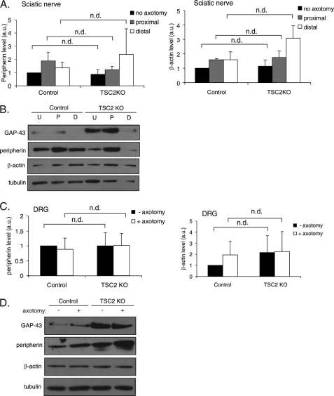
Unlike neurons in the central nervous system (CNS), injured neurons in the peripheral nervous system (PNS) can regenerate their axons and reinnervate their targets. However, functional recovery in the PNS often remains suboptimal, especially in cases of severe damage. The lack of regenerative ability of CNS neurons has been linked to down-regulation of the mTOR (mammalian target of rapamycin) pathway. We report here that PNS dorsal root ganglial neurons (DRGs) activate mTOR following damage and that this activity enhances axonal growth capacity. Furthermore, genetic up-regulation of mTOR activity by deletion of tuberous sclerosis complex 2 (TSC2) in DRGs is sufficient to enhance axonal growth capacity in vitro and in vivo. We further show that mTOR activity is linked to the expression of GAP-43, a crucial component of axonal outgrowth. However, although TSC2 deletion in DRGs facilitates axonal regrowth, it leads to defects in target innervation. Thus, whereas manipulation of mTOR activity could provide new strategies to stimulate nerve regeneration in the PNS, fine control of mTOR activity is required for proper target innervation.


Figures

FIGURE 1.
FIGURE 1.
The mTOR pathway contributes to enhance axonal growth capacity after peripheral nerve injury. Injury to the sciatic nerve of wild-type mice was induced by ligation, and S6 phosphorylation levels were assessed in L4, L5, and L6 DRG cell bodies at the indicated time points. A, time course of S6 phosphorylation in DRG cell bodies showed increased S6 phosphorylation 1 to 2 days following ligation. A representative Western blot is shown for each time point. Cytochrome c (cyt c)was used as loading control. B, quantification of fold change in S6 phosphorylation levels between DRG cell bodies from injured and uninjured nerves shows a 2-fold increase in S6 phosphorylation 1 day after ligation, which reached basal level 4 days following ligation. S6 phosphorylation levels were normalized to loading control. At least four mice were tested for each time point. Data are mean ± S.E. *, p < 0.05 (Student's t test). C, quantification of mean radial projection length of cultured DRGs from wild-type mice treated with rapamycin or dimethyl sulfoxide by intraperitoneal injection and then subjected to sciatic nerve ligation to provoke injury (see “Experimental Procedures” for details). L4, L5, and L6 DRGs were dissected and cultured 4 days later in the presence of NGF. DRGs cultured from injured sciatic nerves showed enhanced axonal outgrowth at 24 h in culture. This effect was blocked partially by rapamycin (n = 4 mice per condition). 100–350 neurons were analyzed per condition. Data are mean ± S.E. *, p < 0.05; **, p < 0.01; ***, p < 0.001 (Student's t test). D, as in C, but a histogram of the distribution in radial projection lengths of injury-conditioned DRGs for one representative experiment. E, selected images of cultured DRGs from C. Images of injury-conditioned DRGs were selected from the far right tail of histograms shown in D. For a comprehensive set of images, see supplemental Fig. 1. Axons were stained with a SMI-31 antibody. Bar, 100 μm.
FIGURE 2.
FIGURE 2.
TSC2KO DRGs display enhanced axonal outgrowth in vitro. A, Western blot of DRG cell bodies and brain lysates from TSC2KO and control animals show that TSC2 protein levels are dramatically reduced in DRGs but not in brain. As expected, S6 phosphorylation level is increased in TSC2KO compared with control animals. A representative Western blot is shown. B, quantification of A (n = 5 mice per genotype). Data are mean ± S.E. **, p < 0.01 (Student's t test). C, quantification of mean radial projection length of naïve and injury-conditioned DRGs cultured from TSC2KO mice and controls in the presence of NGF (see “Experimental Procedures” for details). TSC2KO DRGs show enhanced axonal outgrowth in the absence of a conditioning injury. Injury to the sciatic nerve 4 days prior to dissociation does not further increase axonal outgrowth in TSC2KO DRGs (n = 3 mice per genotype). 60–370 neurons were analyzed per condition. Data are mean ± S.E. n.d., no statistically significant difference. *, p < 0.05; **, p < 0.01 (Student's t test). D, as in C, but a histogram of the distribution in radial projection lengths of cultured naive TSC2KO and control DRGs is shown for one representative experiment. E, selected images of cultured DRGs from C. Images of naive and injury-conditioned TSC2KO DRGs and injury-conditioned control DRGs were selected from the far right tail of their respective radial projection length histogram. A comprehensive set of images is shown in supplemental Fig. 2. Axons were stained with a SMI-31 antibody. F, there was no significant difference in radial projection length between DRGs cultured from wild-type;AdvillinCre/+ mice and those cultured from wild-type;Advillin+/+ mice (n = 3 mice per genotype). 100–200 neurons were measured per genotype. Data are mean ± S.E. n.d., no statistically significant difference (Student's t test). Bar, 100 μm.
FIGURE 3.
FIGURE 3.
TSC2KO DRGs display enhanced regeneration in vivo. TSC2KO and control mice were subject to a sciatic nerve crush, and regeneration of crushed axons was assessed 12 or 24 h later. A, longitudinal section of sciatic nerve dissected 12 or 24 h after crush reveals increased length of GAP-43 positive axons past the crush site in TSC2KO mice compared with control mice for both time points. A dashed line indicates a crush site. B, average GAP-43 intensity at various distances distal to crush sites 12 and 24 h after crush reveal regenerating axons grew longer distances in TSC2KO mice compared with controls. GAP-43 intensity values were normalized to that at the crush site to control for the increased GAP-43 expression level observed from TSC2KO DRGs. Data are mean ± S.E (n = 3 mice per genotype). 3–5 longitudinal sections were analyzed per mouse for both time points. C, regeneration index was measured as the distance away from the crush site in which the average GAP-43 intensity is half that observed at the crush site. TSC2KO mice show a higher regeneration index compared with controls at both 12 and 24 h time points. *, p < 0.05; **, p < 0.01 (Student's t test). Bar, 200 μm.
FIGURE 4.
FIGURE 4.
The mTOR pathway regulates GAP-43 expression. A, naïve TSC2KO DRGs show enhanced cell body and axonal GAP-43 expression in culture. Arrows indicate cell body, and arrowheads indicate axonal tips. B, injury to the sciatic nerve of TSC2KO and control mice was induced by axotomy and GAP-43 levels in nerve portions proximal and distal to the axotomy site and in contralateral noninjured sciatic nerves were analyzed by Western blot 24 h later. Control mice show an increase in GAP-43 expression in sciatic nerve proximal to the injury site. Both the basal level and injury-induced level of GAP-43 expression is enhanced in TSC2KO mice. GAP-43 levels were normalized to loading control (tubulin). Data are mean ± S.E. (n = 3 mice per genotype). *, p < 0.05 (Student's t test). C, representative Western blot from one experiment (one mouse per genotype) from B. D, as in B, but DRG cell bodies were analyzed. GAP-43 levels were increased in DRG cell bodies of TSC2KO mice compared with control. Axotomy did not increase GAP-43 levels in DRG cell bodies in either genotype (n = 3 mice per genotype). **, p < 0.01. (Student's t test). E, representative Western blot from one experiment (one mouse per genotype) from C. F, rapamycin blocks increase in GAP-43 expression after injury. Wild-type mice were injected intraperitoneally with rapamycin or dimethyl sulfoxide vehicle control, and then sciatic nerve injury was induced by ligation. Four days following ligation, unligated and ligated nerve distal and proximal to the ligation site were analyzed for GAP-43 expression. Data are mean ± S.E. (n = 3 mice per condition). *, p < 0.05; *, p < 0.01 (Student's t test). G, representative Western blot from one experiment from F. Bar, 100 μm. a.u., arbitrary units; high exp., high exposure; low exp., low exposure.
FIGURE 5.
FIGURE 5.
TSC2 deletion in DRGs does not affect peripherin or β-actin expression. A, injury to the sciatic nerve of TSC2KO and control mice was induced by axotomy. Peripherin and β-actin levels in nerve portions proximal and distal to the axotomy site and in contralateral noninjured sciatic nerve were analyzed by Western blot 24 h later. No significant difference in basal or injury-induced levels of peripherin or β-actin between TSC2KO and control mice was observed. Peripherin and β-actin levels were normalized to loading control (tubulin). Data are mean ± S.E. (n = 3 mice per genotype). n.d., no statistically significant difference. B, representative Western blot from one experiment (one mouse per genotype) from A. C, as in A, but DRG cell bodies were analyzed. Peripherin and β-actin levels in DRG were not significantly different between TSC2KO and control. Data are mean ± S.E. (n = 3 mice per genotype). D, representative Western blot from one experiment (one mouse per genotype) from C. U, no axotomy; P, proximal; D, distal; a.u., arbitrary units.
FIGURE 6.
FIGURE 6.
TSC2 deletion leads to abnormal target innervation and axon morphology. A, posterior hind paw glaborous skin sections were stained with α-GAP-43 to visualize axonal endings innervating skin. Red dashed lines indicate epidermal layer. Low magnification (15×) confocal images reveal fewer axons penetrating the epidermal layer in TSC2KO mice compared with control. High magnification (60×) confocal images reveal enrichment of endings with excessive branching (arrow) and sharp turning (arrowhead) compared with controls. The presence of Cre recombinase does not contribute to the innvervation defects, as wild-type;AdvillinCre/+ mice did not show significant difference from wild-type;Advillin+/+ littermates or control. Bar, 200 μm for 15× images, 50 μm for 60× images. B, quantification of innervation density reveals loss of skin innervation in TSC2KO mice. Data are mean ± S.E. (n = 3 mice per genotype). 17–56 sections were analyzed per mouse. Quantification of percentage (C) and number (D) of nerve endings with excessive branching (axons with more than two branches at tip or those that turned 90 degrees were counted). *, p < 0.05; n.d., no statistically significant difference (Student's t test). E, cross-section of the peripheral branch of L5 DRG stained with the axonal marker SMI-31 revealed no significant difference in axon number between TSC2 KO and controls. F, quantification of E. (n = 3 mice per genotype). Two sections analyzed per mouse. Bar, 100 μm (E).

References

    1. Case L. C., Tessier-Lavigne M. (2005) Curr. Biol. 15, R749–753 - PubMed
    1. Filbin M. T. (2003) Nat. Rev. Neurosci. 4, 703–713 - PubMed
    1. Yiu G., He Z. (2006) Nat. Rev. Neurosci. 7, 617–627 - PMC - PubMed
    1. Shen Y., Tenney A. P., Busch S. A., Horn K. P., Cuascut F. X., Liu K., He Z., Silver J., Flanagan J. G. (2009) Science 326, 592–596 - PMC - PubMed
    1. Zheng B., Ho C., Li S., Keirstead H., Steward O., Tessier-Lavigne M. (2003) Neuron 38, 213–224 - PubMed

Publication types

MeSH terms

Substances