. 2010 Jul 20;107(29):13165-70.
doi: 10.1073/pnas.1004509107. Epub 2010 Jul 6.

Genetic inhibition of PKA phosphorylation of RyR2 prevents dystrophic cardiomyopathy

Affiliations

Genetic inhibition of PKA phosphorylation of RyR2 prevents dystrophic cardiomyopathy

Satyam Sarma et al. Proc Natl Acad Sci U S A. .

Abstract

Aberrant intracellular Ca(2+) regulation is believed to contribute to the development of cardiomyopathy in Duchenne muscular dystrophy. Here, we tested whether inhibition of protein kinase A (PKA) phosphorylation of ryanodine receptor type 2 (RyR2) prevents dystrophic cardiomyopathy by reducing SR Ca(2+) leak in the mdx mouse model of Duchenne muscular dystrophy. mdx mice were crossed with RyR2-S2808A mice, in which PKA phosphorylation site S2808 on RyR2 is inactivated by alanine substitution. Compared with mdx mice that developed age-dependent heart failure, mdx-S2808A mice exhibited improved fractional shortening and reduced cardiac dilation. Whereas application of isoproterenol severely depressed cardiac contractility and caused 95% mortality in mdx mice, contractility was preserved with only 19% mortality in mdx-S2808A mice. SR Ca(2+) leak was greater in ventricular myocytes from mdx than mdx-S2808A mice. Myocytes from mdx mice had a higher incidence of isoproterenol-induced diastolic Ca(2+) release events than myocytes from mdx-S2808A mice. Thus, inhibition of PKA phosphorylation of RyR2 reduced SR Ca(2+) leak and attenuated cardiomyopathy in mdx mice, suggesting that enhanced PKA phosphorylation of RyR2 at S2808 contributes to abnormal Ca(2+) homeostasis associated with dystrophic cardiomyopathy.


Conflict of interest statement

The authors declare no conflict of interest.

Figures

Fig. 1.
Fig. 1.
Genetic ablation of RyR2-S2808 phosphorylation in mdx mice prevents age-dependent dilated cardiomyopathy. (A) Representative M-mode echocardiogram traces in WT, mdx, and mdx-S2808A mice at 15 mo of age showing reduced contractility and LV dilation in mdx, but not in mdx-S2808A mice. (B) mdx but not mdx-S2808A mice showed a progressive decline of FS (%) compared with WT. (C) Increased EDDs and ESDs is seen in mdx mice at 15 mo of age, which was absent in mdx-S2808A mice. (D) Decline in dP/dt confirmed loss of cardiac contractility at 15 mo of age in mdx but not mdx-S2808A mice. (E) Bar graph shows heart weight–to–body weight ratios 15 mo of age. Numbers in bars indicate number of mice studied per group. *P < 0.05, **P < 0.01, ***P < 0.001.
Fig. 2.
Fig. 2.
Prevention of myocyte disarray and fibrosis in 15-mo-old mdx-S2808A mice. (A) H&E staining of transverse heart sections from 15-mo-old mice showed LV dilation in mdx mice, but not in WT or mdx-S2808A mice. (Scale bar, 0.5 mm.) (B) Masson trichrome staining revealed myocyte disarray and fibrosis in mdx mice (blue fibers) but not in mdx-S2808A mice. (Scale bar, 50 μm.) (C) Quantification of fibrosis in Masson trichrome staining (n = 3 mice in each group). *P < 0.05.
Fig. 3.
Fig. 3.
Development of age-dependent increase in RyR2-S2808 phosphorylation in mdx mice. (A) Representative Western blots and bar graphs with quantification of averaged data showing that phosphorylation of S2808 on RyR2 (RyR2-pS2808) was increased in 15-mo-old mdx mice compared with age-matched WT. The phosphoepitope-specific antibody revealed absence of a signal in RyR2-S2808A mice, as expected. (B) Western blots and bar graphs with averaged data showing that neither total PLN levels nor PKA-phosphorylated PLN (PLN-pS16) levels were significantly altered in mdx mice compared with WT and mdx-S2808A.
Fig. 4.
Fig. 4.
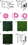
Mutation S2808A in RyR2 protects against ISO-induced acute heart failure and sudden death in mdx mice. (A) Kaplan-Meier survival curves show very low (5%) survival in mdx mice (n = 21) treated with ISO infusion, compared with 100% of WT (n = 13) and 81% of mdx-S2808A mice (n = 21). (B) dP/dt was reduced in mdx mice compared with WT mice, whereas cardiac contractility was significantly improved in mdx-S2808A mice 2 d after ISO infusion. (C) H&E staining of transverse heart sections after 2 d of ISO infusion showed increased RV dilation in mdx mice but not in WT or mdx-S2808A mice. (Scale bar, 1.0 mm.) (DF) WGA staining revealed disruption of sarcolemmal membrane integrity in mdx compared with both WT and mdx-S2808A mice. (Scale bar, 20 μm.) (G) Reduction of membrane integrity evidenced by a lower plasma membrane to nucleus ratio in mdx mice, compared with WT and mdx-S2808A mice (n = 3 mice in each group, 10–20 fields of view analyzed per animal). *P < 0.05, **P < 0.01.
Fig. 5.
Fig. 5.
Genetic inhibition of RyR2-S2808 phosphorylation reduces spontaneous SR Ca2+ releases and SR Ca2+ leak in mdx mice. (A) Representative [Ca2+]i recordings obtained in ventricular myocytes from WT, mdx, or mdx-S2808A mice at the end of a 20-s pacing train (1 Hz), followed by a 40-s pause to check for the occurrence of SR SCRs (marked “v”). SR Ca2+ content was measured by changing superfusate to 10 mM caffeine in 0 Na+, 0 Ca2+ Tyrode. (B) Bar graph shows the percentage of cells exhibiting SCR events during the 20-s pause. SCR occurred more frequently in myocytes from mdx mice, compared with WT and mdx-S2808A myocytes. (C) Bar graph reveals enhanced SR Ca2+ leak normalized to total SR Ca2+ content in mdx mice, compared with WT, and mdx-S2808A. Numbers indicate the number of cells for each condition (four to seven mice per group). *P < 0.05 and **P < 0.01.
Fig. 6.
Fig. 6.
Both pharmacologic and genetic inhibition of PKA phosphorylation of RyR2 prevents SCRs following β-adrenoreceptor activation. (A) Representative [Ca2+]i recording before and after 1 min perfusion of ISO (100 nM). β-Adrenoreceptor activation increased the incidence of SCR events in ventricular myocytes from mdx mice, with PKA inhibitor KT5720 (1 μM) or in mdx-S2808A myocytes. (B) Bar graph shows the percentage of cells exhibiting an increase in the incidence of SCR following ISO perfusion. Numbers indicate the number of cells for each condition (three to four mice per group). *P < 0.05.

References

    1. Petrof BJ, Shrager JB, Stedman HH, Kelly AM, Sweeney HL. Dystrophin protects the sarcolemma from stresses developed during muscle contraction. Proc Natl Acad Sci USA. 1993;90:3710–3714. - PMC - PubMed
    1. Ferlini A, Sewry C, Melis MA, Mateddu A, Muntoni F. X-linked dilated cardiomyopathy and the dystrophin gene. Neuromuscul Disord. 1999;9:339–346. - PubMed
    1. McNally EM. New approaches in the therapy of cardiomyopathy in muscular dystrophy. Annu Rev Med. 2007;58:75–88. - PubMed
    1. Nakayama H, et al. Ca2+- and mitochondrial-dependent cardiomyocyte necrosis as a primary mediator of heart failure. J Clin Invest. 2007;117:2431–2444. - PMC - PubMed
    1. Williams IA, Allen DG. Intracellular calcium handling in ventricular myocytes from mdx mice. Am J Physiol Heart Circ Physiol. 2007;292:H846–H855. - PubMed

Publication types

MeSH terms