. 2010 Jul 20;107(29):13123-8.
doi: 10.1073/pnas.1003825107. Epub 2010 Jul 6.

Correlated memory defects and hippocampal dendritic spine loss after acute stress involve corticotropin-releasing hormone signaling

Affiliations

Correlated memory defects and hippocampal dendritic spine loss after acute stress involve corticotropin-releasing hormone signaling

Yuncai Chen et al. Proc Natl Acad Sci U S A. .

Abstract

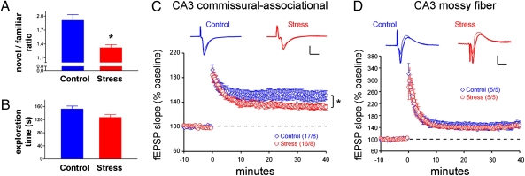
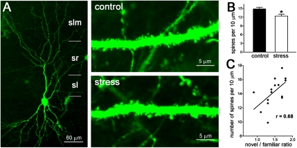
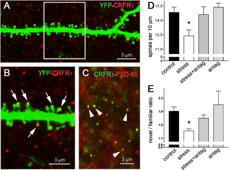
Stress affects the hippocampus, a brain region crucial for memory. In rodents, acute stress may reduce density of dendritic spines, the location of postsynaptic elements of excitatory synapses, and impair long-term potentiation and memory. Steroid stress hormones and neurotransmitters have been implicated in the underlying mechanisms, but the role of corticotropin-releasing hormone (CRH), a hypothalamic hormone also released during stress within hippocampus, has not been elucidated. In addition, the causal relationship of spine loss and memory defects after acute stress is unclear. We used transgenic mice that expressed YFP in hippocampal neurons and found that a 5-h stress resulted in profound loss of learning and memory. This deficit was associated with selective disruption of long-term potentiation and of dendritic spine integrity in commissural/associational pathways of hippocampal area CA3. The degree of memory deficit in individual mice correlated significantly with the reduced density of area CA3 apical dendritic spines in the same mice. Moreover, administration of the CRH receptor type 1 (CRFR(1)) blocker NBI 30775 directly into the brain prevented the stress-induced spine loss and restored the stress-impaired cognitive functions. We conclude that acute, hours-long stress impairs learning and memory via mechanisms that disrupt the integrity of hippocampal dendritic spines. In addition, establishing the contribution of hippocampal CRH-CRFR(1) signaling to these processes highlights the complexity of the orchestrated mechanisms by which stress impacts hippocampal structure and function.


Conflict of interest statement

The authors declare no conflict of interest.

Figures

Fig. 1.
Fig. 1.
Short multimodal stress impairs learning and memory and area CA3 synaptic plasticity in select dendritic lamina. (A) Mice experiencing a 5-h combined psychological/physical stress failed to distinguish an object that they had seen before from a novel one. Control mice explored the novel object preferentially, whereas stressed mice spent almost equal time exploring an object they had encountered 6 h earlier and a novel one, suggesting they did not distinguish between them. *P < 0.05. (B) Stressed mice that were allowed a 90-min recovery period explored two objects for the same duration as control mice, eliminating the possibility that their apparent memory defects were a result of freezing or poor motivation. n = 10 mice per group. (C) Slices prepared from mice < 1 h after multimodal stress (red) exhibited deficient LTP magnitude at area CA3 C/A synapses as compared with controls (blue). Representative traces from baseline (thin lines) and 30-min postinduction (thick lines) are shown. Values in parentheses indicate numbers of slices. *P = 0.01 for minutes 30–40. (D) Mossy fiber potentiation tested in slices from the same animals as in C showed no effect of stress. Results are shown as means ± SEM. Values in parentheses indicate number of mice. [Scale bars in C and D: 1 mV (vertical bar) and 10 ms (horizontal bar).]
Fig. 2.
Fig. 2.
Reduced spine density after short multimodal stress and its correlation to cognitive function. (A) (Left) An area CA3 hippocampal neuron expressing YFP. The apical dendritic arbor is delineated, showing its span within stratum lucidum (sl), stratum radiatum (sr), and stratum lacunosum-moleculare (slm). Stress-vulnerable dendritic spines are located in the stratum radiatum. (Right) Segments of apical dendrites (within the stratum radiatum) from a control mouse (Upper) and from a mouse killed after a 5-h multimodal stress (Lower) are shown. (B) The reduced spine density is quantified; *P < 0.05. (C) Correlation between spine density in apical dendrites (third and fourth branches in the stratum radiatum) and the ratio of time spent exploring a novel vs. a familiar object in individual mice. r = 0.68 (Spearman's correlation coefficient); P = 0.0055.
Fig. 3.
Fig. 3.
Blocking the effects of short multimodal stress on dendritic spines abrogates cognitive deficits. (A) The CRH receptor CRFR1 resides on dendritic spine heads, as shown by confocal microscopy of dendrites from YFP-expressing mice. (B) Boxed area in A; arrows denote spine heads expression the CRH receptor. (C) Confocal microscopic image obtained after dual immunohistochemistry for the receptor and PSD-95 (arrowheads point to dual-labeled puncta typical of spine heads). The receptor colocalizes with PSD-95. (D) Mean spine density in area CA3 stratum radiatum segments of YFP-labeled pyramidal neurons in control mice, mice subjected to stress, mice subjected to stress with intracerebral pretreatment with the CRFR1 antagonist NBI 30775 (15 μg in 1 μL) (stress + antag), and control mice administered an equal dose of NBI 30775 (antag). All mice had indwelling cannulae and received vehicle or NBI 30775 to eliminate potential confounders of the surgical or infusion procedures. n = 6–7 mice per group. (E) Novel object recognition ratio in mice subjected to the four treatments described in D. Stressed mice explored the novel object significantly less than the other groups. n = 6–7 mice per group. Asterisks in D and E indicate significant difference from control values.

References

    1. Squire LR, Wixted JT, Clark RE. Recognition memory and the medial temporal lobe: A new perspective. Nat Rev Neurosci. 2007;8:872–883. - PMC - PubMed
    1. Eichenbaum H, Yonelinas AP, Ranganath C. The medial temporal lobe and recognition memory. Annu Rev Neurosci. 2007;30:123–152. - PMC - PubMed
    1. Andersen P, Moser E, Moser MB, Trommald M. Cellular correlates to spatial learning in the rat hippocampus. J Physiol Paris. 1996;90:349. - PubMed
    1. Morris RG, et al. Elements of a neurobiological theory of the hippocampus: The role of activity-dependent synaptic plasticity in memory. Philos Trans R Soc Lond B Biol Sci. 2003;358:773–786. - PMC - PubMed
    1. Larson J, Lynch G. Induction of synaptic potentiation in hippocampus by patterned stimulation involves two events. Science. 1986;232:985–988. - PubMed

Publication types

Substances