. 2010 Jul 13;107(28):12704-9.
doi: 10.1073/pnas.0910106107. Epub 2010 Jul 6.

Therapeutic potential of appropriately evaluated safe-induced pluripotent stem cells for spinal cord injury

Affiliations

Therapeutic potential of appropriately evaluated safe-induced pluripotent stem cells for spinal cord injury

Osahiko Tsuji et al. Proc Natl Acad Sci U S A. .

Abstract

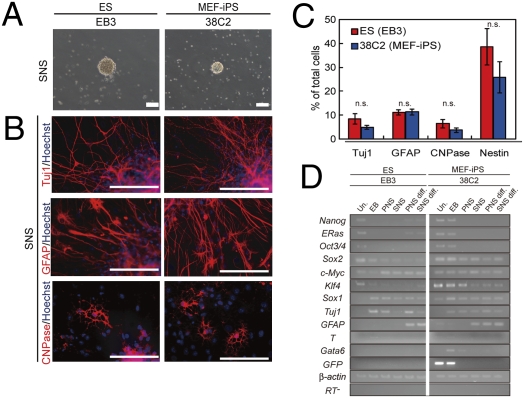
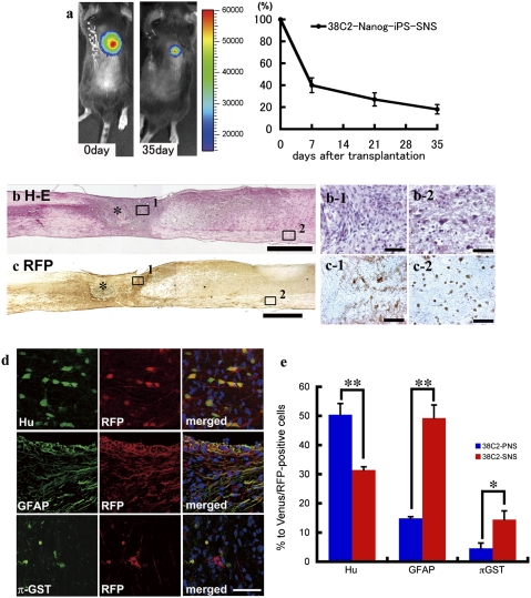
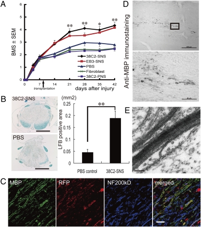
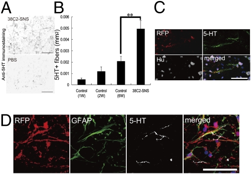
Various types of induced pluripotent stem (iPS) cells have been established by different methods, and each type exhibits different biological properties. Before iPS cell-based clinical applications can be initiated, detailed evaluations of the cells, including their differentiation potentials and tumorigenic activities in different contexts, should be investigated to establish their safety and effectiveness for cell transplantation therapies. Here we show the directed neural differentiation of murine iPS cells and examine their therapeutic potential in a mouse spinal cord injury (SCI) model. "Safe" iPS-derived neurospheres, which had been pre-evaluated as nontumorigenic by their transplantation into nonobese diabetic/severe combined immunodeficiency (NOD/SCID) mouse brain, produced electrophysiologically functional neurons, astrocytes, and oligodendrocytes in vitro. Furthermore, when the safe iPS-derived neurospheres were transplanted into the spinal cord 9 d after contusive injury, they differentiated into all three neural lineages without forming teratomas or other tumors. They also participated in remyelination and induced the axonal regrowth of host 5HT(+) serotonergic fibers, promoting locomotor function recovery. However, the transplantation of iPS-derived neurospheres pre-evaluated as "unsafe" showed robust teratoma formation and sudden locomotor functional loss after functional recovery in the SCI model. These findings suggest that pre-evaluated safe iPS clone-derived neural stem/progenitor cells may be a promising cell source for transplantation therapy for SCI.


Conflict of interest statement

The authors declare no conflict of interest.

Figures

Fig. 1.
Fig. 1.
Neural differentiation of pre-evaluated safe MEF-iPS cells in vitro. (A) Neurospheres derived from EB3 ES cells and 38C2 iPS cells. (Scale bar: 200 μm.) (B) Immunocytochemical analysis of neural cell marker proteins in the differentiated SNSs derived from EB3 ES and 38C2 iPS cells. (Scale bar: 100 μm.) (C) Neural differentiation efficiencies of neurospheres derived from EB3 ES and 38C2 iPS cells. (n = 5, n.s.). (D) RT-PCR analysis of undifferentiated cells (Un.), EBs, PNSs, SNSs, differentiated PNSs (PNS diff.), and SNSs (SNS diff.) of the EB3-ES and 38C2 iPS cells.
Fig. 2.
Fig. 2.
Transplanted SNSs derived from safe MEF-iPS clones survive without any evidence of tumorigenesis and differentiate into trilineage neural cells in the injured spinal cord. (A) Representative BLI images of a mouse in which CBRluc-expressing 38C2 iPS-SNSs were transplanted into the injured spinal cord (Left, immediately after transplantation; Right, 42 d after transplantation). Quantification of the photon intensity revealed that ≈60% of the grafted cells were lost within 7 d after transplantation, and ≈20% of the cells survived 35 d after transplantation. Values are means ± SEM (n = 6). (B) H&E and (C) anti-RFP DAB staining of sagittal sections of the spinal cord 42 d after injury (38C2 iPS-SNS transplanted). There was no evidence of tumorigenesis (B). No significant nuclear atypia was observed in magnified images of the boxed areas showing the lesion epicenter (B-1) or white matter caudal to the transplantation site (B-2). Grafted cells survived and were diffusely distributed rostral and caudal to the lesion site (C). Higher-magnification images of the boxed areas showing the lesion site (C-1) and white matter caudal to the lesion site (C-2). *Lesion epicenter. (D) Immunohistochemical analyses of 38C2 iPS-SNSs grafted into spinal cord 42 d after injury, revealing grafted cells double-positive for RFP and markers of neural lineages. (E) Quantitative analyses of Hu+ neurons, GFAP+ astrocytes, and π-GST+ oligodendrocytes. Values are means ± SEM (n = 3 each; *P < 0.05, **P < 0.01).
Fig. 3.
Fig. 3.
SNS derived from a safe MEF-iPS clone differentiate into mature oligodendrocytes and promote remyelination. (A) Time course of functional recovery of hind limbs evaluated by BMS. 38C2 iPS-SNS, n = 19; EB3 ES-SNS, n = 15; PBS, n = 12; adult fibroblasts, n = 13; 38C2 iPS-PNS, n = 13. *P < 0.05, **P < 0.01. (B) LFB staining of axial sections of the spinal cord at the lesion epicenter 42 d after injury; 38C2 iPS-SNS–transplanted (Upper Left) and PBS control (Lower Left) animals. Quantification of LFB-positive areas at the lesion epicenter 42 d after injury (Right, n = 7 each; **P < 0.01). (C) Immunohistochemistry of 38C2 iPS-SNS–derived mature oligodendrocytes (MBP+). Grafted cells were integrated into myelin sheath. (D) Anti-MBP DAB staining of sagittally sectioned spinal cord of a shiverer mouse 8 wk after transplantation. MBP+ myelin was detected in the area caudal to the lesion epicenter. (Lower) Higher-magnification image of the boxed area. (E) EM pictures of the injured spinal cord of a 38C2 iPS-SNS–grafted shiverer mouse exhibiting a prominent major dense line and intraperiod lines in multiple compacted lamellae. (Scale bars: B, 500 μm; D Upper, 200 μm; C and D Lower, 50 μm; and E, 0.1 μm.)
Fig. 4.
Fig. 4.
SNSs derived from a safe MEF-iPS clone promote serotonergic innervation of the dorsal cord and result in better functional recovery of the hindlimbs. (A) 38C2 iPS-SNS transplantation promoted the growth of 5HT+ serotonergic fibers in the distal spinal cord. Axial sections of 38C2 iPS-SNS–transplanted (Upper) and PBS control mice (Lower). (B) Quantitative analysis of 5HT+ serotonergic fibers of distal cord in the PBS control (1, 2, and 6 wk postinjury) and 38C2 iPS-SNS transplantation groups (6 wk postinjury; 1 and 2 wk postinjury, n = 3 each; 6 wk postinjury and 38C2 SNS, n = 7 each; **P < 0.01). (C and D) Immunohistochemistry of 38C2 iPS-SNS–derived neurons (C, RFP+, Hu+) and astrocytes (D, RFP+, GFAP+) closely associated with 5HT+ serotonergic fibers. (Scale bars: A, 100 μm; C, 20 μm; D, 50 μm.)
Fig. 5.
Fig. 5.
Characterization and transplantation of SNSs derived from safe and unsafe TTF-iPS cells. (A) Neurospheres derived from 1A2 ES cells, 335D1, 256H13, and 256H18 iPS cells. (Scale bar: 200 μm.) (B) The differentiation potential of TTF-iPS-derived SNSs tested in vitro by immunocytochemical analyses of neural cell markers; Tuj1 for neurons, GFAP for astrocytes, and CNPase for oligodendrocytes. (Scale bar: 100 μm.) (C) Time course of functional recovery of the hindlimbs evaluated by BMS. 335D1 iPS-SNS: n = 9 each; 256H13 and 256H18 iPS-SNS: n = 9; 1A2 ES-SNS: n = 9; PBS control: n = 8. *P < 0.05, **P < 0.01. (D–F) H&E sagittal sections of the spinal cord 42 d after injury. (D) 335D1 iPS-SNS, (E) 256H18 iPS-SNS, and (F) 256H13 iPS-SNS grafted mice. There was no evidence of tumorigenesis in the 335D1 iPS-SNS grafted mice (D), whereas teratoma formation was detected within the injured spinal cord in both 256H18 iPS-SNS (E), and 256H13 iPS-SNS (F) grafted mice. (G) Anti-Nanog DAB staining of sagittally sectioned spinal cord of 256H18 and 256H13 iPS-SNS–transplanted animals 35 d after transplantation.

References

    1. Björklund A, Lindvall O. Cell replacement therapies for central nervous system disorders. Nat Neurosci. 2000;3:537–544. - PubMed
    1. Okano H. Stem cell biology of the central nervous system. J Neurosci Res. 2002;69:698–707. - PubMed
    1. Lindvall O, Kokaia Z, Martinez-Serrano A. Stem cell therapy for human neurodegenerative disorders—how to make it work. Nat Med. 2004;10(Suppl):S42–S50. - PubMed
    1. Martino G, Pluchino S. The therapeutic potential of neural stem cells. Nat Rev Neurosci. 2006;7:395–406. - PubMed
    1. Lindvall O, Kokaia Z. Stem cells for the treatment of neurological disorders. Nature. 2006;441:1094–1096. - PubMed

Publication types

MeSH terms